Method and storage medium for contrast enhanced ultrasound quantization imaging

A technology of contrast and ultrasound, which is applied in the direction of ultrasound/sonic/infrasonic equipment control, ultrasound/sonic/infrasonic diagnosis, application, etc. It can solve the problems that cannot be displayed, cannot be used to display organ structures, cannot display different blood vessel structures, etc.

Pending Publication Date: 2022-04-22
SHENZHEN MINDRAY BIO MEDICAL ELECTRONICS CO LTD
View PDF0 Cites 1 Cited by
  • Summary
  • Abstract
  • Description
  • Claims
  • Application Information

AI Technical Summary

Problems solved by technology

However, slow low-volume blood flow, for example in capillaries, may not be visualized by conventional Doppler methods, and thus Doppler ultrasound may not be able to visualize different vascular structures (e.g., different sized vessels) in the region of interest. Blood vessel)
Also, for example, Doppler ultrasound cannot be used to visualize organ structures to visualize blood flow distribution and perfusion in regions of interest

Method used


Image

  • Method and storage medium for contrast enhanced ultrasound quantization imaging
  • Method and storage medium for contrast enhanced ultrasound quantization imaging
  • Method and storage medium for contrast enhanced ultrasound quantization imaging

Examples


Embodiment Construction

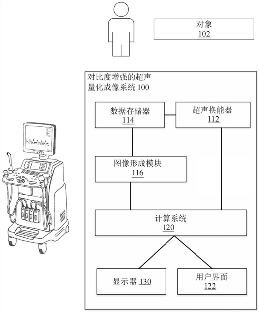
[0026] figure 1 An exemplary environment for a contrast-enhanced quantified ultrasound imaging (CEUS QI) system 100 for contrast-enhanced quantified ultrasound imaging is shown in accordance with various embodiments. CEUS QI system 100 may include additional components, or fewer components, or alternative components, depending on implementation needs.

[0027] In some embodiments, CEUS QI system 100 may include ultrasound transducer 112, data storage 114, image formation module 116, computing system 120, user interface 122, and display 130, wherein one or more of the foregoing components are optional. Ultrasound transducer 112 can be coupled to data storage 114 and computing system 120, data storage 114 can be coupled to image forming module 116, image forming module 116 can be coupled to computing system 120, computing system 120 can be coupled to display 130 and user interface 122 . Any coupled modules may transmit signals between each other. Ultrasound transducer 112, da...


PUM


Abstract

A method and computer readable storage medium for contrast enhanced ultrasound quantization imaging. One of the methods comprises: for each position in a region of interest, acquiring a time-varying ultrasonic signal associated with the region of interest within a time period; for each location, a processed time-varying ultrasound signal is determined based at least on the acquired time-varying ultrasound signal, where: a portion where a local increase exceeds a threshold is mapped, and a portion where a local increase does not exceed the threshold is flattened; an image sequence of the region of interest is generated based at least on the processed time-varying ultrasound signals at each location, where the generated image sequence displays signal variations of the processed time-varying ultrasound signals in one or more structures in the region of interest to represent the one or more structures.

Description

technical field [0001] The present application relates generally to contrast-enhanced ultrasound quantitative imaging, and more particularly to methods and systems for visualizing the direction and distribution of blood flow in vascular structures. Background technique [0002] Ultrasound imaging is a non-invasive medical testing technique that examines soft tissues in the body, such as blood vessels, to reveal structural details of internal tissues and fluid flow. Many ultrasound imaging systems utilize injectable ultrasound contrast agents to obtain high-contrast images, which are desirable for visualizing blood flow in internal organ structures. For example, images of the direction and distribution of blood flow often aid doctors in diagnosis and treatment. [0003] Doppler ultrasound is an imaging test that shows blood flowing through blood vessels and is commonly used to detect and image fluid flow in the body, such as blood flow. In Doppler ultrasound, the frequency ...

Claims


Application Information

Patent Timeline
Patent Type & Authority Applications(China)
IPC IPC(8): A61B8/06A61B8/00
CPCA61B8/06A61B8/481A61B8/469A61B8/488A61B8/461A61B8/54A61B8/5223
Inventor 吉挺澜陆兆龄
Owner SHENZHEN MINDRAY BIO MEDICAL ELECTRONICS CO LTD
Why Eureka
Try Eureka