Method for preparing liver organoid from peripheral blood mononuclear cells

An organoid and liver technology, applied in the field of preparation of liver organoids from peripheral blood mononuclear cells, can solve the problems of worsening liver function, limited access to liver tissue, and increased bleeding risk.

Pending Publication Date: 2022-05-31
SUN YAT SEN UNIV
View PDF7 Cites 3 Cited by
  • Summary
  • Abstract
  • Description
  • Claims
  • Application Information

AI Technical Summary

Problems solved by technology

[0006] One is primary hepatic stem cells obtained from human liver tissue by isolation and culture, but obtaining liver tissue is an invasive operation, especially for patients with abnormal liver function or failure, invasive operations such as liver puncture or liver resection will Significantly increased risk of bleeding or worsening liver function, so access to liver tissue is limited and difficult

Method used


Image

  • Method for preparing liver organoid from peripheral blood mononuclear cells
  • Method for preparing liver organoid from peripheral blood mononuclear cells
  • Method for preparing liver organoid from peripheral blood mononuclear cells

Examples


Embodiment 1

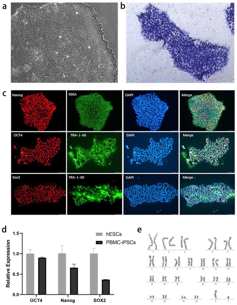
[0091] Example 1 Induction of PBMCs into iPSCs with Multiple Differentiation Potentials

[0092] In this example, PBMCs were isolated and extracted from the peripheral blood of the donor (the collection of the specimen was approved by the ethics committee of the hospital).

[0093] 1. Experimental method

[0094] 1. Extraction of PBMCs by Ficoll-Paque gradient centrifugation

[0095] Use heparin anticoagulant tubes to extract 3ml of peripheral blood from the donor, and place it on ice for later use. Add 6ml of lymphocyte separation medium to a 15ml centrifuge tube, and place the tube at an angle of 45°. After mixing the anticoagulant blood with a pipette tip, slowly add it over the lymphocyte separation solution along the wall of the test tube, so that the lymphocyte separation solution and the anticoagulant blood form a clear dividing line. Centrifuge the centrifuge tube at 12 degrees Celsius, 2500rpm, increase speed to 1, decrease speed to 0, and centrifuge for 20 minutes...

Embodiment 2

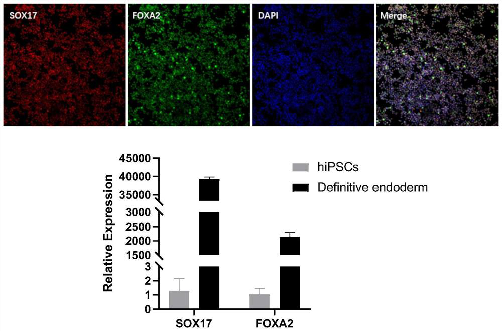
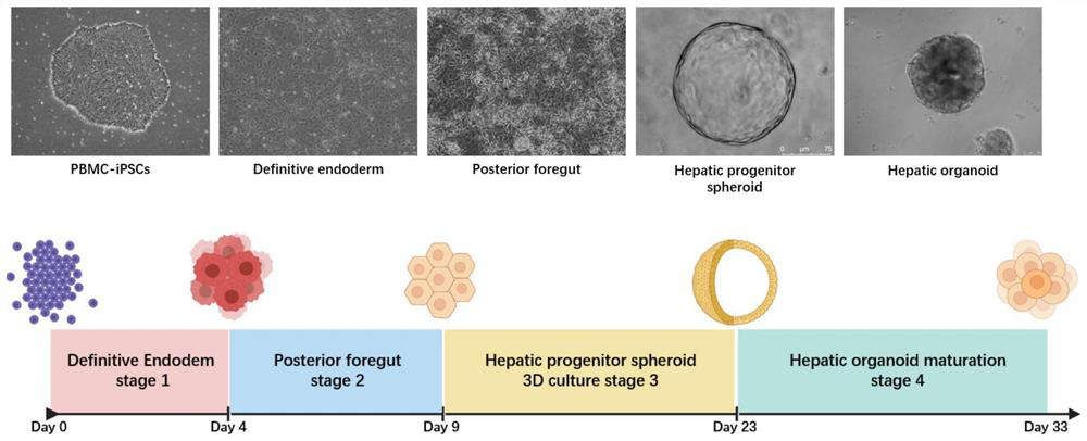
[0116] Example 2 Inducing iPSCs with multilineage differentiation potential into terminal foregut endoderm cells (Posterior Foregut) through committed endoderm cells

[0117] 1. Experimental method

[0118] The iPSCs cells with multilineage differentiation potential prepared in the above Example 1 were incubated with mTeSR medium at 37°C and 5% CO 2 Under the condition of culture, the culture medium was changed every 2 days to expand iPSCs. When the cell density reached 80%, it was digested with Accutase for 5 minutes, culture medium was added to stop the digestion, and centrifuged at 1100 rpm for 4 minutes. Cell count, according to 0.5~1×10 5 piece / cm 2 Inoculated in cell plates and cultured overnight using mTeSH+10μM Y27632 for the next step of endoderm induction.

[0119] Afterwards, on the first day (D1), replace endoderm induction medium 1: RPMI 1640 medium + 1% (v / v) B27 + 50ng / ml WNT3A protein + 100ng / ml Activin A protein, culture for one day, second to third Day (...

Embodiment 3

[0124] Example 3 Resuspension culture of terminal foregut endoderm cells in Matrigel to induce hepatic progenitor spheres

[0125] 1. Experimental method

[0126] 1. Terminal foregut endoderm cells were resuspended in Matrigel

[0127] On the ninth day (D9), the flat-cultured terminal foregut endoderm cells were washed twice with PBS, digested into single cells by adding Accutace, and then culture medium was added to terminate the digestion, collected in a centrifuge tube, centrifuged at 1100 rpm for 4 minutes. Count the cells, collect 200,000 to 400,000 cells in a 1.5ml centrifuge tube at a cell density of 10,000 to 20,000 cells / 50 μl Matrigel, discard the supernatant after centrifugation, and place the centrifuge tube on ice for later use.

[0128] Take 1ml of Matrigel matrix collagen solution and put it on ice to melt into a liquid state, and place the pipette tip in a refrigerator at 4°C to pre-cool. Add 1ml of Matrigel into the centrifuge tube containing the cell pellet...


PUM


Abstract

The invention discloses a method for preparing liver organoid from peripheral blood mononuclear cells (PBMCs). A liver organoid model which has individual genetic information and fully simulates liver development and pathological and physiological processes of liver-related diseases in vitro is obtained in a relatively noninvasive manner. According to the method, the collected PBMCs are reprogrammed into induced pluripotent stem cells (iPSCs), the iPSCs are further induced and differentiated into the liver organs, the target is achieved through the two steps, the iPSCs can be infinitely amplified and stored in vitro, and a basis is provided for large-scale production of the liver organs.

Description

technical field [0001] The invention relates to the technical field of stem cells, in particular to a method for preparing liver organoids from peripheral blood mononuclear cells. Background technique [0002] The liver is the largest metabolic organ in the human body. There are a wide variety of liver-based genetic metabolic diseases, including fatty liver, congenital biliary atresia, citrin deficiency, α-1 antitrypsin deficiency, tyrosinemia type Ⅰ, liver lenticular degeneration and so on. The research on the occurrence and development of these liver diseases requires good in vivo and in vitro models. Although various animal models have been constructed at home and abroad according to various diseases, there are great differences between animals and humans in terms of development, physiological structure and pathological state. Animal models It is difficult to replicate the progression of human disease, so better disease models are urgently needed. [0003] At this time,...

Claims


Application Information

Patent Timeline
Patent Type & Authority Applications(China)
IPC IPC(8): C12N5/071C12N5/074
CPCC12N5/0672C12N5/0671C12N2500/30C12N2500/46C12N2500/32C12N2500/90C12N2501/345C12N2501/16C12N2501/30C12N2501/415C12N2501/11C12N2501/998C12N2501/12C12N2501/237C12N2500/40C12N2500/38C12N2506/45C12N2509/10C12N2509/00
Inventor 项鹏柯琼范明明李伟强
Owner SUN YAT SEN UNIV
Why Eureka
Try Eureka