
Ultrasonic image processing method and equipment

An ultrasonic image and area technology, applied in the field of medical image processing, can solve the problems of complex operation and low efficiency, and achieve the effect of simplifying operation and improving work efficiency.

Pending Publication Date: 2022-07-05
SHENZHEN MINDRAY BIO MEDICAL ELECTRONICS CO LTD
View PDF0 Cites 2 Cited by
  • Summary
  • Abstract
  • Description
  • Claims
  • Application Information

AI Technical Summary

Problems solved by technology

[0004] The embodiment of the present invention provides an ultrasonic image processing method and equipment, which are used to solve the problems of complicated operation and low efficiency existing in the existing fetal cardiothoracic ratio measurement process

Method used


Image

  • Ultrasonic image processing method and equipment
  • Ultrasonic image processing method and equipment
  • Ultrasonic image processing method and equipment

Examples


Embodiment Construction

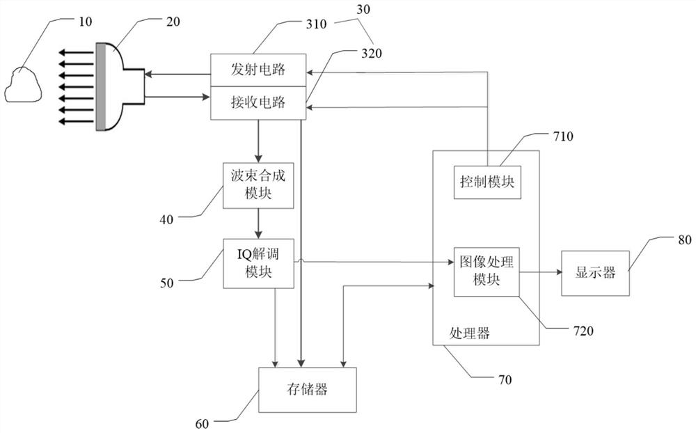
[0068] The present invention will be further described in detail below through specific embodiments in conjunction with the accompanying drawings. Wherein similar elements in different embodiments have used associated similar element numbers. In the following embodiments, many details are described so that the present application can be better understood. However, those skilled in the art will readily recognize that some of the features may be omitted under different circumstances, or may be replaced by other elements, materials, and methods. In some cases, some operations related to the present application are not shown or described in the specification, in order to avoid the core part of the present application being overwhelmed by excessive descriptions, and for those skilled in the art, these are described in detail. The relevant operations are not necessary, and they can fully understand the relevant operations according to the descriptions in the specification and gener...


PUM


Abstract

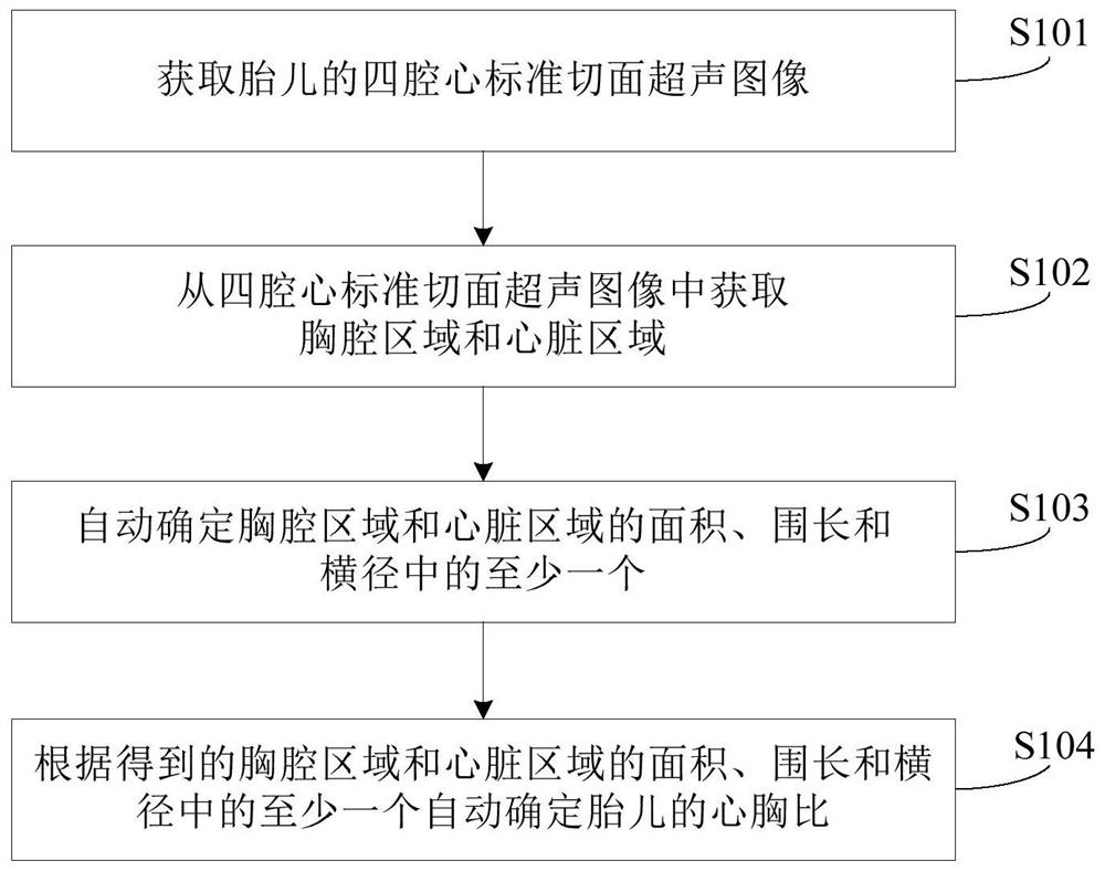
The embodiment of the invention provides an ultrasonic image processing method and equipment. The method comprises the following steps: acquiring a four-cavity heart standard section ultrasonic image of a fetus; obtaining a thoracic cavity area and a heart area from the four-cavity heart standard section ultrasonic image; automatically determining at least one of an area of the thoracic cavity region and the heart region, a girth of the thoracic cavity region and the heart region, and a transverse diameter of the thoracic cavity region and the heart region; a cardiothoracic ratio of the fetus is automatically determined based on at least one of an area of the thoracic and cardiac regions, a girth of the thoracic and cardiac regions, and a transverse diameter of the thoracic and cardiac regions. According to the method provided by the embodiment of the invention, the automatic measurement of the heart-chest ratio of the fetus is realized based on the ultrasonic image of the fetus, the operation is simplified, and the working efficiency of a clinician is improved.

Description

technical field [0001] The invention relates to the technical field of medical image processing, in particular to an ultrasonic image processing method and device. Background technique [0002] In clinical practice, ultrasound technology is often used to dynamically observe fetal growth and development, fetal activity, fetal heartbeat, breathing and swallowing. Among the many ultrasound examination items during pregnancy, especially in the second and third trimesters, the measurement of fetal cardiothoracic ratio helps to screen the fetus for cardiac malformation or other congenital heart disease, which is very important for the assessment of fetal growth and development. Clinical attention is increasing day by day. [0003] In actual clinical measurements, doctors need to manually trace the heart region and the thoracic region in the obtained fetal ultrasound images, so as to calculate the cardiothoracic ratio. The above process is easily affected by the clinical experien...

Claims


Application Information

Patent Timeline
IPC IPC(8): A61B8/08A61B8/00G06T7/00G06T7/11G06T7/62
CPCA61B8/0866A61B8/085A61B8/5215A61B8/483G06T7/0012G06T7/11G06T7/62A61B2503/02G06T2207/30048G06T2207/10136
Inventor 刘超越邹耀贤林穆清陈卫程
Owner SHENZHEN MINDRAY BIO MEDICAL ELECTRONICS CO LTD
Learn More