Preparation method of non-squamous epithelial cell simulant
A technology of squamous epithelium and mimics, which is applied in the field of analysis and detection, and can solve the problems of poor test stability of non-squamous epithelial cells
- Summary
- Abstract
- Description
- Claims
- Application Information
AI Technical Summary
Problems solved by technology
Method used
Image
Examples
preparation example Construction
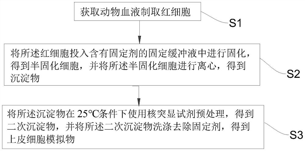
[0032] see Figure 1 to Figure 11 , the present invention provides a method for preparing a non-squamous epithelial cell simulant, comprising the following steps: .
[0033] S1 obtains animal blood to prepare red blood cells;
[0034] Specifically, the blood of the animal is selected from the blood of amphibians, such as frogs and turtles.
[0035] Specific way:
[0036] S11 obtains amphibian blood, and centrifuges the blood for stratification to obtain a stratified liquid;
[0037] S12 removes the supernatant and the white blood cell layer of the layered liquid to obtain a red blood cell layer;
[0038] In S13, the red blood cell layer was washed by centrifugation with normal saline to obtain the red blood cells.
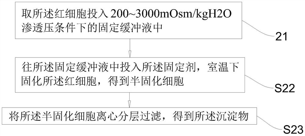
[0039] S2 put the red blood cells into a fixation buffer containing a fixative for solidification to obtain semi-solidified cells, and centrifuge the semi-solidified cells to obtain a precipitate;
[0040] Specifically, the fixative is an aldehyde fixative sel...
Embodiment 1
[0050] S001 filter fresh anticoagulated bullfrog blood;
[0051] S002 Centrifuge the anticoagulated bullfrog blood at 1500 r / min for 5 min, remove the supernatant and white blood cells, and obtain pure red blood cells;
[0052] S003 wash the red blood cells by centrifugation with normal saline;
[0053] S004 takes the precipitate in step 3, resuspends the cells with fixation buffer containing 0.1% formaldehyde, and solidifies at room temperature for 24h;
[0054] S005 Centrifuge the semi-solidified cells at 1500 r / min for 5 min, and discard the supernatant;
[0055] S006 was added to the fixation buffer containing 0.1% formaldehyde with nuclear highlighting reagent containing 1% acetic acid, the cells were resuspended with this solution, and the cells were solidified at 25°C for 24h;
[0056] S007 was washed with PBS buffer to remove fixative.
[0057] The prepared simulant was tested in a urine visible component analyzer based on the principle of flow microscopy, and the res...
Embodiment 2
[0059] S011 filter fresh anticoagulated bullfrog blood;
[0060] S012 Centrifuge the anticoagulated bullfrog blood at 1500 r / min for 5 min, remove the supernatant and white blood cells, and obtain pure red blood cells;
[0061] S013 wash the red blood cells by centrifugation with normal saline;
[0062] S014 takes the precipitate in step 3, resuspends the cells with fixation buffer containing 0.02% glutaraldehyde, and solidifies at room temperature for 24h;
[0063] S015 Centrifuge the semi-solidified cells at 1500 r / min for 5 min, and discard the supernatant.
[0064] S016 was added with 1% acetic acid-containing nucleophore reagent in fixation buffer containing 0.02% glutaraldehyde, and the cells were resuspended with this solution, and solidified at 25°C for 24h;
[0065] S017 was washed with PBS buffer to remove fixative.
[0066] The prepared simulant was tested in a urine visible component analyzer based on the principle of flow microscopy, and the results were as fol...
PUM
 Abstract
Description
Claims
Application Information
 - R&D
- Intellectual Property
- Life Sciences
- Materials
- Tech Scout
- Unparalleled Data Quality
- Higher Quality Content
- 60% Fewer Hallucinations
Browse by: Latest US Patents, China's latest patents, Technical Efficacy Thesaurus, Application Domain, Technology Topic, Popular Technical Reports.
© 2025 PatSnap. All rights reserved.Legal|Privacy policy|Modern Slavery Act Transparency Statement|Sitemap|About US| Contact US: help@patsnap.com


