Blood brain barrier in-vitro model and preparation method thereof

A blood-brain barrier and model technology, applied in biochemical equipment and methods, artificial cell constructs, enzymes, etc., to achieve effects that are beneficial to drug development and screening

Pending Publication Date: 2022-07-29
SHENZHEN UNIV
View PDF4 Cites 0 Cited by
  • Summary
  • Abstract
  • Description
  • Claims
  • Application Information

AI Technical Summary

Problems solved by technology

[0004] The purpose of this application is to provide an in vitro model of the blood-brain barrier and its preparation method, aiming to solve the problem of how to construct an in vitro model of the blood-brain barrier that is more similar to the real blood-brain barrier structure

Method used


Image

  • Blood brain barrier in-vitro model and preparation method thereof
  • Blood brain barrier in-vitro model and preparation method thereof
  • Blood brain barrier in-vitro model and preparation method thereof

Examples


preparation example Construction

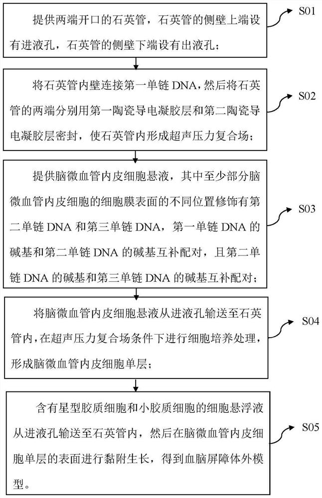
[0025] The first aspect of the embodiments of the present application provides a method for preparing an in vitro model of the blood-brain barrier, such as figure 1 shown, including the following steps:

[0026] S01: provide a quartz tube with openings at both ends, the upper end of the side wall of the quartz tube is provided with a liquid inlet hole, and the lower end of the side wall of the quartz tube is provided with a liquid outlet hole;

[0027] S02: connect the inner wall of the quartz tube with the first single-stranded DNA, then seal both ends of the quartz tube with the first ceramic conductive gel layer and the second ceramic conductive gel layer respectively, so that an ultrasonic pressure composite field is formed in the quartz tube;

[0028] S03: Provide a suspension of cerebral microvascular endothelial cells. Different positions on the cell membrane surface of at least part of the cerebral microvascular endothelial cells in the suspension of cerebral microvasc...

Embodiment 1

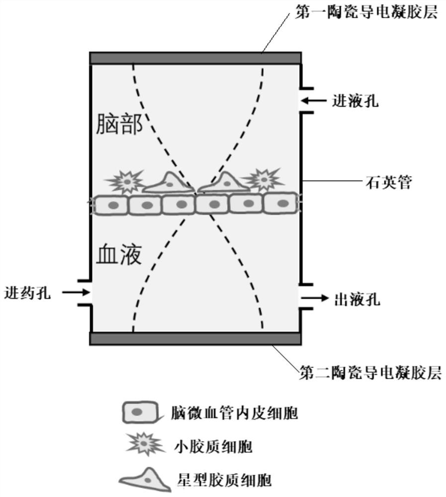
[0058] An in vitro model of the blood-brain barrier, such as figure 2 As shown, it includes a quartz tube and a first ceramic conductive gel layer and a second ceramic conductive gel layer respectively located at both ends of the quartz tube. The quartz tube is provided with a monolayer of cerebral microvascular endothelial cells formed under the condition of an ultrasonic pressure compound field. The cells in the microvascular endothelial cell monolayer and between the brain microvascular endothelial cell monolayer and the inner wall of the quartz tube are connected by DNA chains, and astrocytes and microglia adhere and grow on the upper surface of the brain microvascular endothelial cell monolayer. Plasma cells.

[0059] The preparation method of the blood-brain barrier in vitro model comprises the following steps:

[0060] S11: Pre-customized with quartz material such as figure 1 In the quartz tube channel structure shown, the upper end of the side wall of the quartz tub...


PUM

PropertyMeasurementUnit
The inside diameter ofaaaaaaaaaa

Abstract

The invention belongs to the technical field of tissue engineering simulation, and particularly relates to a blood-brain barrier in-vitro model and a preparation method thereof. The blood-brain barrier in-vitro model comprises a quartz tube, and a first ceramic conductive gel layer and a second ceramic conductive gel layer which are respectively positioned at two ends of the quartz tube, and a brain microvascular endothelial cell monolayer formed under the condition of an ultrasonic pressure composite field is arranged in the quartz tube. Cells in the brain microvascular endothelial cell monolayer are connected through DNA chains, the brain microvascular endothelial cell monolayer and the inner wall of the quartz tube are connected through DNA chains, and astrocytes and microglia grow on the upper surface of the brain microvascular endothelial cell monolayer in an adhesion mode. The blood-brain barrier in-vitro model can simulate a blood-brain barrier structure more truly, and direct co-culture contact among different cells can reflect actual conditions more truly, so that the blood-brain barrier in-vitro model can be beneficial to drug development and screening of a central nervous system and has a good application prospect.

Description

technical field [0001] The present application belongs to the technical field of tissue engineering simulation, and in particular relates to an in vitro model of the blood-brain barrier and a preparation method thereof. Background technique [0002] The blood-brain barrier (BBB), also known as the cerebrovascular barrier or the blood-brain barrier, refers to a "barrier" between the blood vessels and the brain that selectively prevents certain substances from entering the brain from the blood, such as brain capillaries. The barriers between blood vessel walls and glial cells between plasma and brain cells, and between plasma and cerebrospinal fluid, formed by the choroid plexus, prevent certain (mostly harmful) substances from entering the brain from the bloodstream. [0003] The blood-brain barrier is a typical feature of the human central nervous system. It is a dynamic interface between the blood circulatory system and the central nervous system (CNS) of the brain. This de...

Claims


Application Information

Patent Timeline
IPC IPC(8): C12N5/071C12N5/079C12N13/00C12Q1/02
CPCC12N5/0697C12N5/069C12N13/00G01N33/5058G01N33/5064C12N2502/28C12N2502/086C12N2533/10C12N2533/14C12N2527/00C12N2503/02G01N2500/10
Inventor 许太林罗勇张学记
Owner SHENZHEN UNIV
Why Eureka
Try Eureka