

Method for detecting neoplastic disorders in a solubilized body sample
一种样本中、样本的技术,应用在检测癌症,的体外诊断装置,检测分子标记,抑制因子水平的检测试剂盒领域,能够解决没有公开LBC样本制备方法等问题
- Summary
- Abstract
- Description
- Claims
- Application Information
AI Technical Summary
Problems solved by technology
Method used
Image

Examples
Embodiment 1
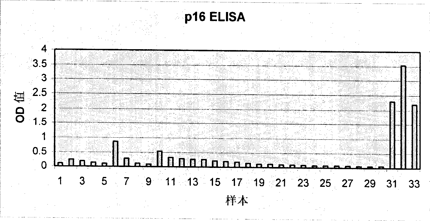
[0166] Example 1: Detecting cervical intraepithelial neoplasia by ELISA
[0167] Provided 33 cervical swabs present in the lysis medium, the cyclin-dependent kinase inhibitor p16 in the solution prepared from the cells contained in the swab was detected by an ELISA-based method INK4a The overexpression. The ELISA test is implemented as follows:
[0169] Put the cervical swab brush into a 15 ml container containing 2 ml of mtm lysis medium (2% Triton-X 100 in PBS, 0.4% SDS, 0.6 mM PMSF). Cervical cells present on the brush are lysed for at least 20 hours. Then transfer the lysate of the cervical swab sample into a 2ml test tube, and centrifuge at 4°C (28.000×g for 15 minutes (16.600rpm high-speed centrifuge JEC Multi RF)); transfer the supernatant to a new test tube. The supernatant can be stored at -20°C.
[0170] (B) Implement ELISA
[0171] Coated ELISA plate
[0172] Dilute p16 with PBS INK4a The specific antibody clones mtm E6H4 stock solution to a usab...
Embodiment 2
[0203] Example 2: Detection of intraepithelial neoplasia of the cervix by lateral flow detection
[0204] Perform routine PAP testing on 9 provided cervical swabs in PreservCyt (Cytyc Corporation, Boxborough, MA) solution, while using a lateral flow-based method to detect cyclin in a solution prepared from the cell suspension obtained from the swab Inhibitor of dependent kinase p16 INK4a The overexpression. The flow detection is implemented as follows:
[0205] (A) Cell lysis
[0206] From PreservCyt TMThe 10ml cell suspension of the individual's cervical swab sample provided by the fixed material was transferred into a 15ml reaction vessel. The sample was centrifuged at 1500×g at room temperature for 15 minutes (3000rpm, Heraeus Varifuge, rotor8074); the supernatant was discarded and the remaining methanol was allowed to evaporate (room temperature, 15 minutes); the precipitate was dissolved in 500μl of lysis buffer, and then transferred to 1.5 ml reaction solution container. The...
Embodiment 3
[0219] Example 3: Detection of p16 using RT-PCR method INK4a And p14 ARF Transcription of
[0220] Cervical samples from 50 individuals were used for this analysis. For each individual, obtain two samples, one in the Universal Collection Medium (Universal Collection Medium) and the other in the PreservCyt TM In solution. Two samples were obtained in the same testing process. For each individual, based on the data from PreservCyt TM Diagnosis is obtained by analysis of thin cervical specimens prepared in the solution. In this study, 20 samples were selected to be diagnosed as NILM, 20 samples were diagnosed as LSIL, and 10 samples were diagnosed as HSIL. According to the following method, using RT-PCR method to analyze the p16 of all samples at the mRNA level INK4a And p14 ARF The transcription level is determined:
[0221] To perform the analysis, cells were removed from UCM and PreservCyt by centrifugation TM Precipitation in solution. The obtained precipitate is directly subject...
PUM
 Abstract
Description
Claims
Application Information