

Methods and reagents for the rapid and efficient isolation of circulating cancer cells
A cell-based, reagent-based technology with application in the field of oncology and diagnostic assays that addresses issues of limited sensitivity and practical, limited, and insignificant improvement in the ability to count targeted cells or examine them
- Summary
- Abstract
- Description
- Claims
- Application Information
AI Technical Summary
Problems solved by technology
Method used
Image


Examples
Embodiment 1
[0104] Formulating improved magnetic nanoparticles for efficient isolation of rare cells from whole blood
[0105] Rare cells (eg, tumor cells in patients with epithelially derived tumors, fetal cells in maternal blood, etc.) may be present at a frequency of less than one rare cell per ml of blood. The number of blood smears required to detect these rare cells is surprisingly large. Assume that there are 10 rare cells per 10ml of blood, corresponding to 5-10×10 7 If there are 10 rare cells in 1 white blood cell, the cells can be transferred to a microscope slide by cytocentrifugation or sedimentation, stained with an antibody specific for the rare cells of interest, and then read manually or automatically. The maximum number of cells that can be transferred to one slide is about 500,000 cells, which means that 100-200 slides are required to process 10ml of blood. The time required for analysis by this method makes the method impractical and economically unfeasible. Therefor...
Embodiment 2
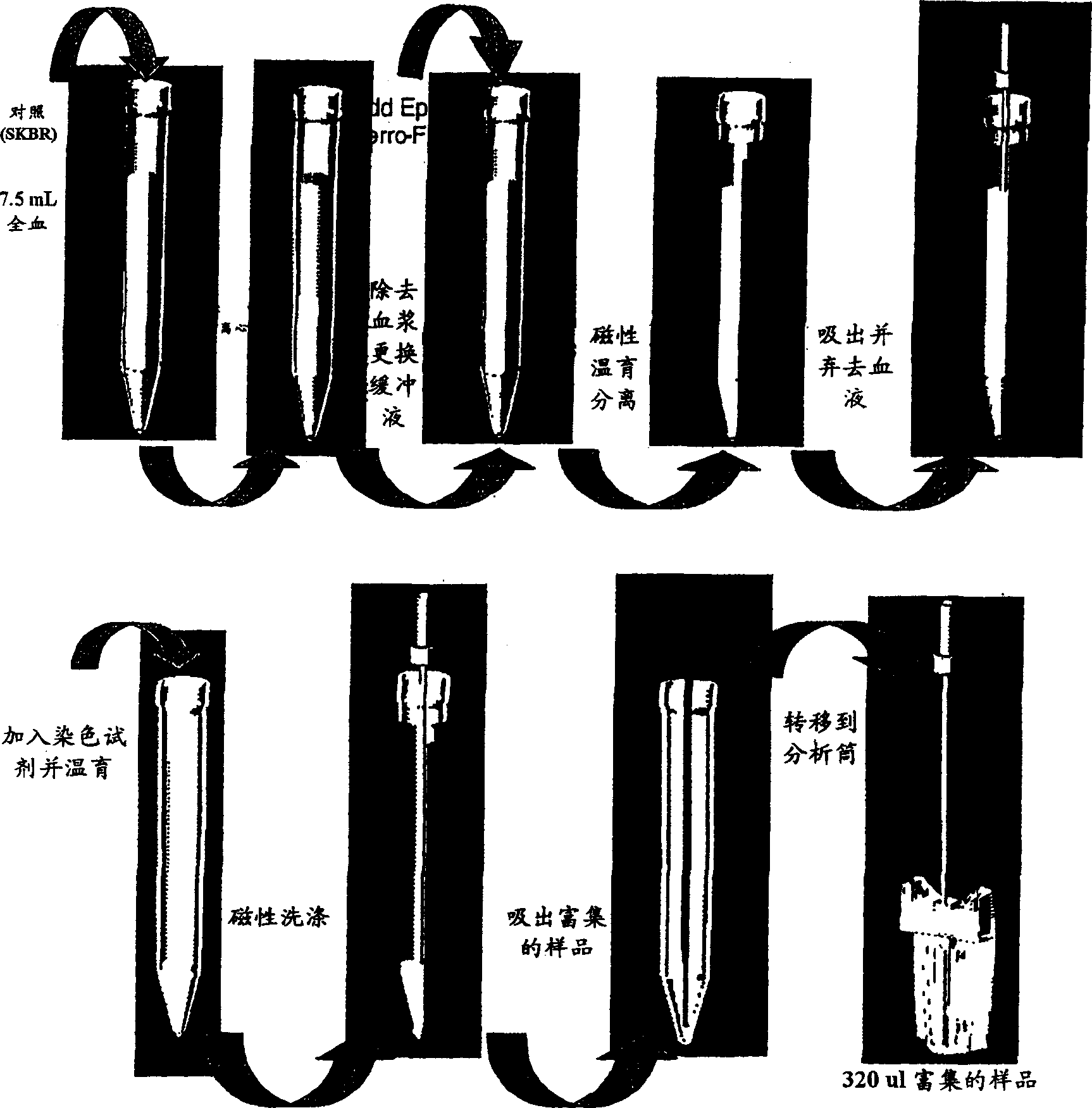
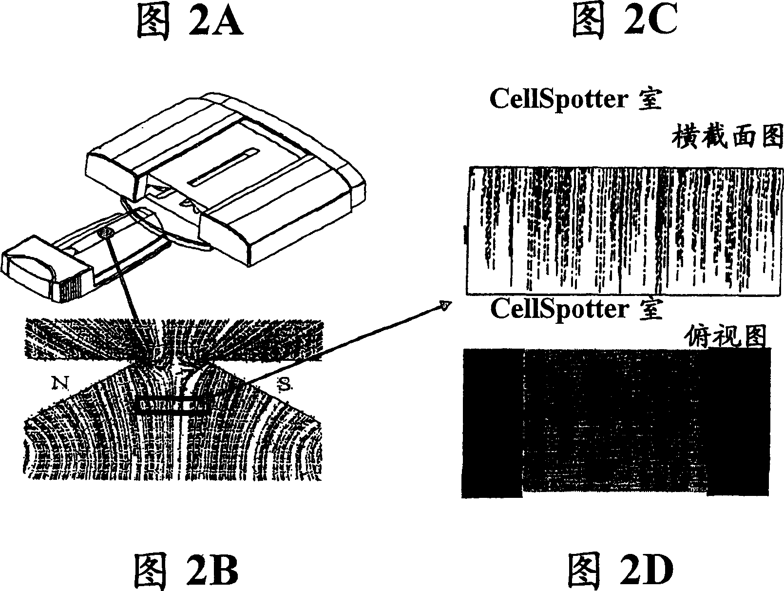
[0115] Semi-automated sample preparation and analysis to identify circulating tumor cells
[0116] A semi-automated system was developed to process and analyze 7.5 ml of blood for the presence of epithelial-derived tumor cells. Immunomagnetic labeling and isolation of cells of epithelial origin in blood. Differentially fluorescently labeled cells captured magnetically and placed in the analysis chamber. Differentiate debris, hematopoietic cells, and circulating tumor cells (CTCs) of epithelial origin using four-color fluorescence imaging. An algorithm is applied on the captured images to calculate internal controls and identify all objects likely to be classified as tumor cells based on size and immunophenotype. A small image of each object is presented on the user interface, from which the user can determine the presence of tumor cells. The internal control showed consistent and reproducible results across different systems and operators when processing blood from norm...
Embodiment 3
[0146] Counting circulating epithelial cells in metastatic breast cancer patients
[0147] The following methods are provided to facilitate the implementation of the following embodiments.
[0148] patient. After obtaining consent from the participants, 8-20 ml blood samples were taken from controls as well as breast, prostate and colon cancer patients. Blood was drawn from some of these patients at several time points over the course of a year. Blood samples were drawn into Vacutainer tubes (Becton-Dickinson) containing EDTA as an anticoagulant. The samples were stored at room temperature and processed within 24 hours of collection. Circulating epithelial cells were counted in peripheral blood samples from breast cancer patients, prostate cancer patients, and colon cancer patients, and in normal controls without malignancy. Get diagnostic data, therapeutic intervention, and clinical status from patient tables. The institutional review board of the partner institution...
PUM
 Abstract
Description
Claims
Application Information