Process of separating endothelial ancestral cell from human fat tissue

An endothelial progenitor cell and separation method technology is applied in the field of obtaining seed cells, and achieves the effects of low cost, wide source of raw materials and easy operation

Active Publication Date: 2007-03-21
SHANGHAI TISSUE ENG LIFE SCI
View PDF0 Cites 8 Cited by
  • Summary
  • Abstract
  • Description
  • Claims
  • Application Information

AI Technical Summary

Problems solved by technology

But using FN to separate EPC from adipose tissue has not been reported at home and abroad.

Method used


Image

  • Process of separating endothelial ancestral cell from human fat tissue
  • Process of separating endothelial ancestral cell from human fat tissue
  • Process of separating endothelial ancestral cell from human fat tissue

Examples


Embodiment 1

[0042] Method for isolating endothelial progenitor cells from human adipose tissue

[0043] (1) Human adipose tissue was digested with 0.01% type II collagenase for 30 minutes;

[0044] (2) Centrifuge at 1200g for 5 minutes;

[0045] (3) with 5×10 4 piece / cm 2 Inoculate on a fibronectin (FN)-coated culture dish, cultivate with DMEM medium containing 2% fetal bovine serum, and change medium for 6 hours;

[0046] (4) When the cells are fused to 80-90%, subculture, the subculture density is 2×10 4 piece / cm 2 , after in vitro amplification to the second generation, induction is carried out.

Embodiment 2

[0048] cell induction

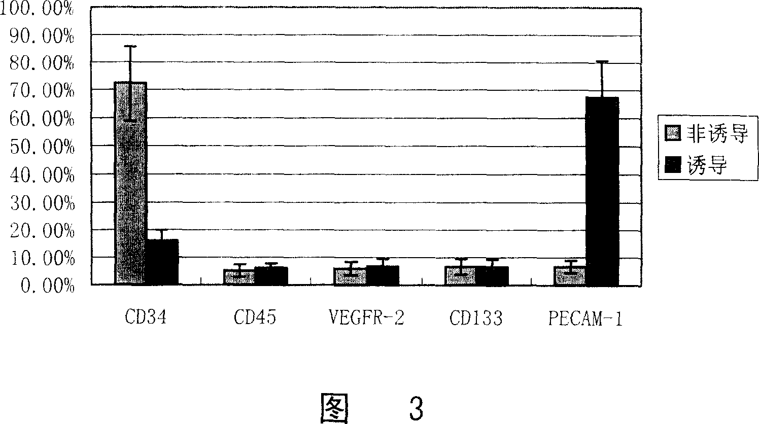
[0049] The second-generation cells obtained by the method described in Example 1 were divided into induction group and non-induction group, and the non-induction group was cultured with DMEM medium containing 2% fetal bovine serum; the induction group was cultured with 2% fetal bovine serum and VEGF, Culture in EGM-2 medium with growth factors such as bFGF.

Embodiment 3

[0051] detection

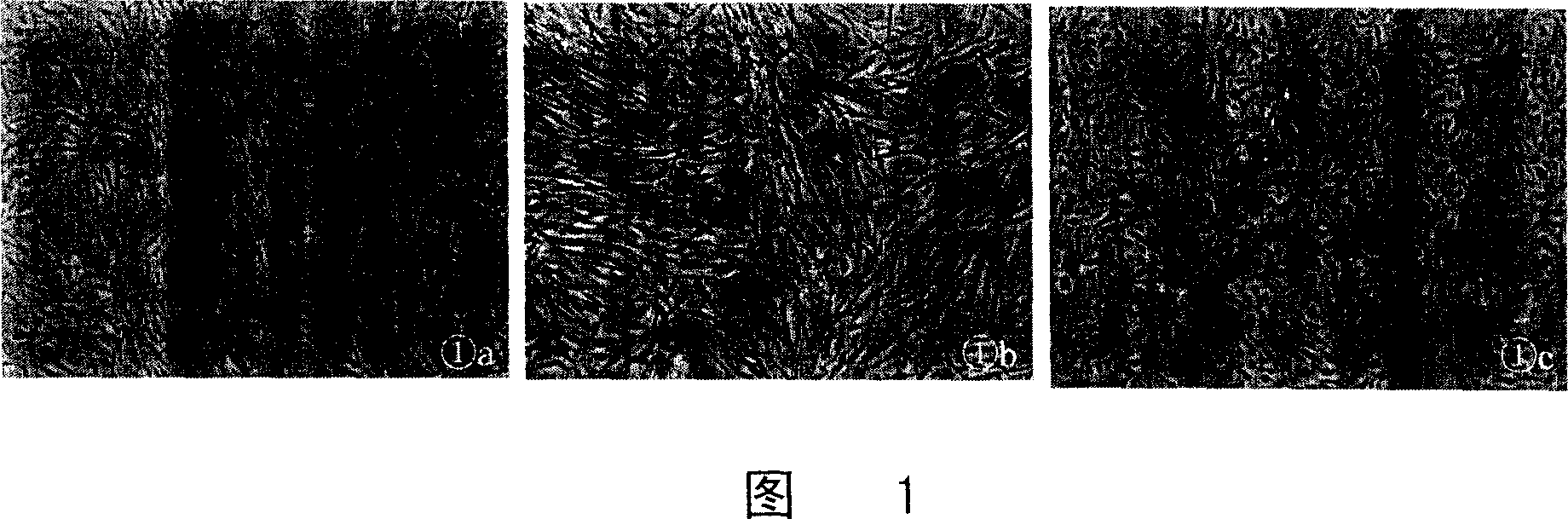
[0052] 1. Observation of cell morphology

[0053] The results are shown in Figure 1. After 12 days of induction, the cells showed a typical endothelial cell "paving stone" structure. The primary cells were spindle-shaped cells (Figure 1a); the second-generation cells in the non-induced group were still spindle-shaped (Figure 1b), while the cells in the induced group showed a typical "paving stone"-like arrangement (Figure 1c).

[0054]Normal endothelial cells have a "paving stone" like morphology. We transformed the received "spindle-shaped" cells into an endothelial-like morphology under appropriate culture conditions. Morphologically, the "spindle" cells transformed into a normal endothelial cell-like morphology, suggesting that the isolated "spindle" cells are precursors (ie, progenitors) of endothelial cells.

[0055] 2. Cell immunofluorescence detection:

[0056] After culturing and induction for 12 days respectively, fix the cells with ethanol:acet...


PUM


Abstract

The process of separating endothelial ancestral cell from human fat tissue relates to method of obtaining seed cell in tissue engineering. Morphological change and cellular immunofluorescene show that the induced cell possesses the phenotype of mature endothelial cell and function of ingesting low density lipoprotein. The detection result with flow cytometer shows that the inducing system possesses very high inducing efficiency. The induced cell forms dendrite bifurcation structure during 3D culture.

Description

technical field [0001] The invention relates to a method for obtaining seed cells in tissue engineering. Background technique [0002] At present, the endothelial seed cells required for the construction of tissue-engineered blood vessels are mainly derived from mature endothelial cells of large vessels or microvessels. The following reasons limit their application: first, the source of donor blood vessels is limited; The capacity is limited and cannot meet the requirements of tissue engineering for the number of seed cells. [0003] As early as 1997, Asahara T et al. found that there were precursor cells capable of differentiating into vascular endothelial cells in postnatal circulating peripheral blood. Considering their continuation relationship with angioblasts in embryonic development, they were called vascular endothelial progenitor cells (endothelial progenitor cells). cell, EPC). EPC is a kind of precursor cells that can circulate, proliferate and differentiate int...

Claims


Application Information

Patent Timeline
Patent Type & Authority Applications(China)
IPC IPC(8): C12N5/08C12N5/0775
Inventor 崔磊曹谊林刘伟刘波
Owner SHANGHAI TISSUE ENG LIFE SCI
Why Eureka
Try Eureka