
Ultrasonic diagnostic contrast imaging at moderate MI levels

A technology of ultrasonic diagnosis and imaging system, which is applied in the field of medical diagnostic imaging system, and can solve problems such as nonlinear contrast agent signal damage

Inactive Publication Date: 2007-06-06
KONINKLIJKE PHILIPS ELECTRONICS NV
View PDF1 Cites 3 Cited by
  • Summary
  • Abstract
  • Description
  • Claims
  • Application Information

AI Technical Summary

Problems solved by technology

But when using a more modest MI above 0.1 with a more rigid contrast agent, for example on the order of 0.3-0.4, the non-linear contrast agent signal is compromised, with unacceptable levels of harmonics returning from the tissue

Method used


Image

  • Ultrasonic diagnostic contrast imaging at moderate MI levels
  • Ultrasonic diagnostic contrast imaging at moderate MI levels
  • Ultrasonic diagnostic contrast imaging at moderate MI levels

Examples


Embodiment Construction

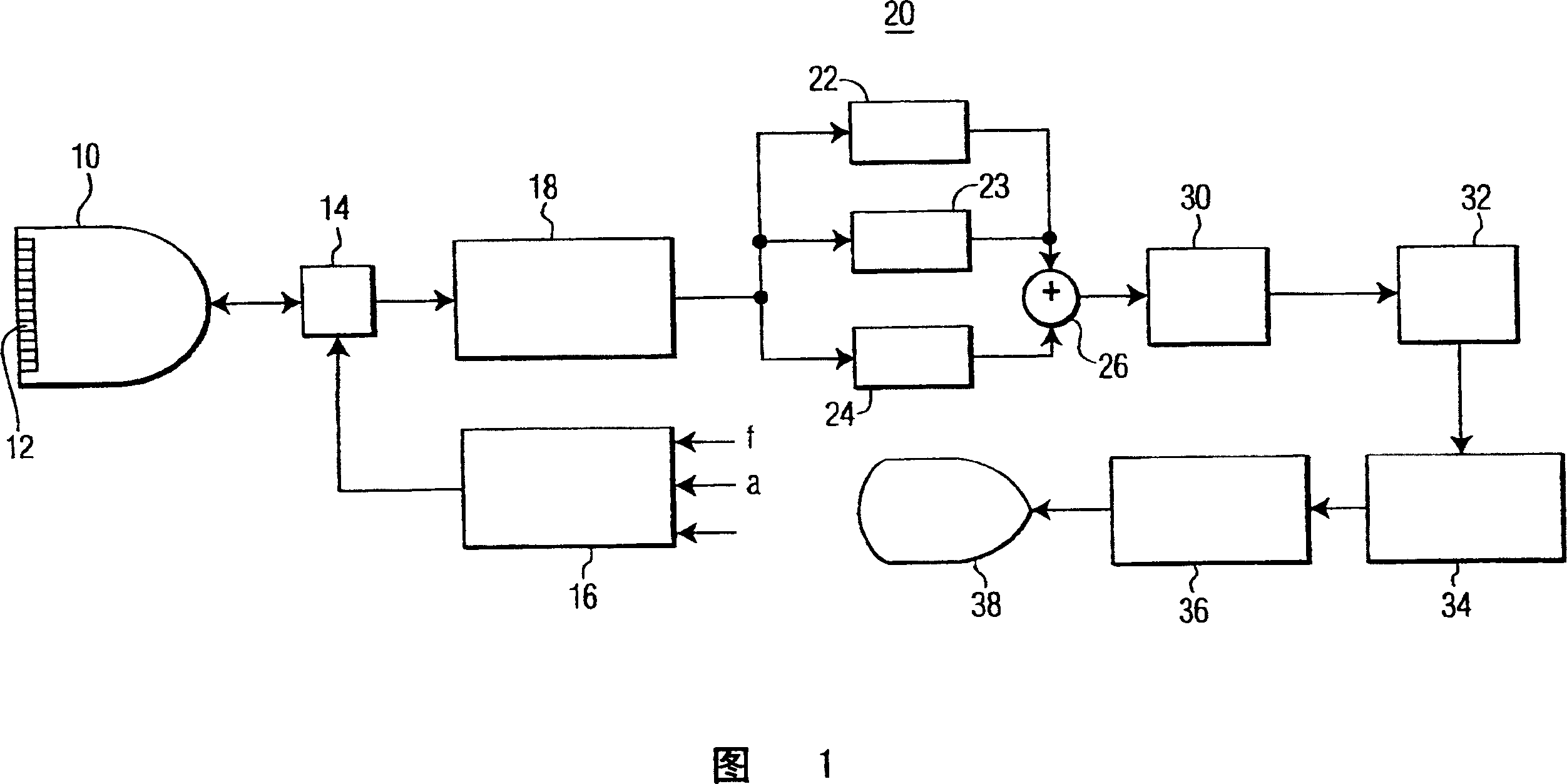
[0012] Referring first to Fig. 1, there is shown an ultrasonic diagnostic imaging system constructed according to the principles of the present invention. The ultrasound system of FIG. 1 utilizes a transmitter 16 which transmits a multi-pulse sequence for generating an echo signal with a nonlinear response. The transmitter is coupled to the elements of the array transducer 12 of the scan head 10 through the transmit / receive switch 14. The transmitter responds to a number of control parameters that modulate the characteristics of the transmitted pulse. The transmitter can control the transmission frequency f of the pulse wave and / or the amplitude a of the pulse. The transmitter can also control the relative phase of the pulse wave. This modulation allows the echoes received in response to these pulses to be combined to separate non-linear echo signal components for imaging.

[0013] In FIG. 1, the transducer array 12 receives echoes from the human body containing linear and non-lin...


PUM


Abstract

A method and device for imaging contrast agents which oscillate nonlinearly in a nondestructive mode at an MI in excess of 0.1 is described. Three transmit pulses are transmitted in each beam direction which are differently modulated. In an illustrated embodiment the transmit pulses are symmetrically differently phase modulated at 0 degrees, 120 degrees and 240 degrees. The echoes received in response to each transmit pulse are stored and combined by a pulse inversion processor. Pulse inversion processing results in separation of the third harmonic to the relative exclusion of the first and second harmonic signal components. Third harmonic images of the contrast image are formed which exhibit a relatively low tissue background.

Description

Technical field [0001] The invention relates to a medical ultrasound imaging system, in particular to a medical diagnostic imaging system that uses a contrast agent and uses moderate mechanical index emission waves. Background technique [0002] The use of ultrasound contrast agents can significantly enhance ultrasound imaging of blood flow. The microbubbles of the contrast agent can be designed to oscillate non-linearly or rupture when ultrasonic waves penetrate. Such oscillation or rupture will make the echo returning from the microbubbles rich in nonlinear components. The echo is received, and the non-linear component is separated from the echo returned by the tissue through filtering or a two-pulse separation technique called pulse inversion. The images generated from these echoes can clearly segment the blood flow and vasculature containing the contrast agent. [0003] Contrast agents are usually imaged using high mechanical index (MI) energy or low MI energy. When imaging w...

Claims


Application Information

Patent Timeline
IPC IPC(8): A61B8/00G01S7/52
CPCG01S7/52039G01S15/8959G01S7/52038G01S15/8963G01S7/52A61B8/14
Inventor M·布鲁斯M·阿弗基欧
Owner KONINKLIJKE PHILIPS ELECTRONICS NV
Learn More