Method estimating a pseudo Hounsfield Unit value

a pseudo-housfield value and unit value technology, applied in the field of magnetic resonance imaging, can solve the problems of not being able to cover the anatomical variations between patients, not being able to generate estimated electron density maps from magnetic resonance images, and not being able to achieve strong correlation of intensity values for training

Active Publication Date: 2018-10-30
KONINKLJIJKE PHILIPS NV
View PDF8 Cites 0 Cited by
  • Summary
  • Abstract
  • Description
  • Claims
  • Application Information

AI Technical Summary

Benefits of technology

[0029]This embodiment may have the advantage that the estimation of pseudo CT Hounsfield Unit (HU) values from magnetic resonance images.
[0030]By determinination of a weighted contribution of different tissue classes pseudo HU values for different tissue classes can be easily combined. Signal intensities inside a volume element for different tissue classes (retrieved from different MR images) can simply be compared with each other to determine the tissue classes' individual contributions to the pseudo HU value.
[0031]Using MR imaging it is complicated to directly determine a pseudo Hounsfield value for certain types of tissue classes as some tissue classes do not show signal on an MR image. By substracting relative prevalence of a first and / or second tissue class from a total tissue prevalence also a relative prevalence for low signal tissue classes can be obtained. In turn this information can be used to calculate a pseudo Hounsfield unit value for a volume element comprising a low signal tissue class. Thereby, the proposed method improves the estimation of pseudo CT Hounsfield Unit (HU) values from magnetic resonance images.

Problems solved by technology

Creating estimated electron density maps from magnetic resonance (MR) images is a problem that occurs e.g. with integrated PET / MR systems, where an attenuation maps needs to be estimated, or with radiation therapy planning based on MR, where electron density maps are required for the treatment simulation.
However, due to the physics of the image acquisition, MR intensities do not uniquely correspond to electron density, hence those maps cannot be derived from the MR image by a simple lookup operation, as is commonly done when estimating these maps from CT images.
However, in highly variable anatomical regions like the pelvic region, registration may not be able to cover the anatomical variations between patients, e.g. bladder / bowel filling or movement, resection of structures (e.g. kidneys, liver parts) or pathologic changes.
Further, partial volume effects are ignored, which is especially problematic for thin structures, e.g. the cortical bone.
However, this is a definite drawback of the method, since only tissue-mixture types represented by a separate Gaussian can be assigned reliably.
This may be achievable in the head, where there is little anatomical variation between acquisitions, however, for highly variable regions, such as the pelvis, this strong correlation of intensity values for training may not be achievable.

Method used


Image

  • Method estimating a pseudo Hounsfield Unit value
  • Method estimating a pseudo Hounsfield Unit value
  • Method estimating a pseudo Hounsfield Unit value

Examples


Embodiment Construction

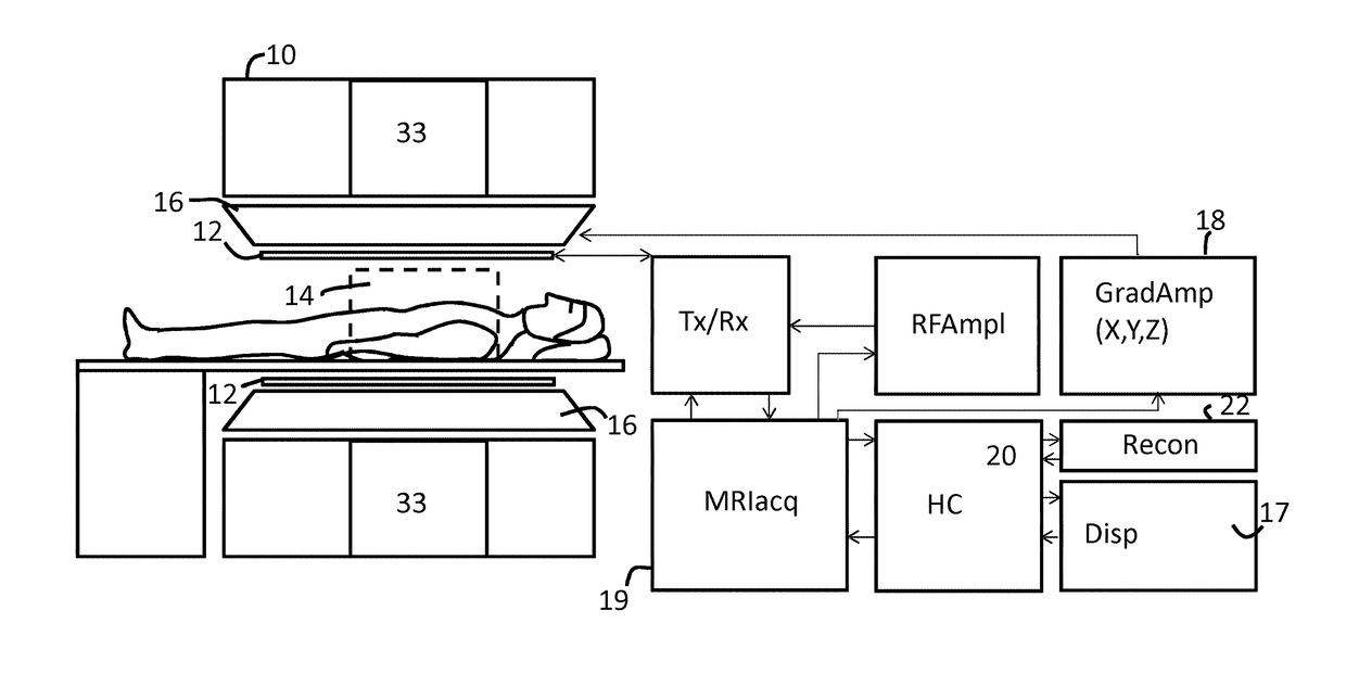
[0049]Like numbered elements in these figures are either equivalent elements or perform the same function. Elements which have been discussed previously will not necessarily be discussed in later figures if the function is equivalent.

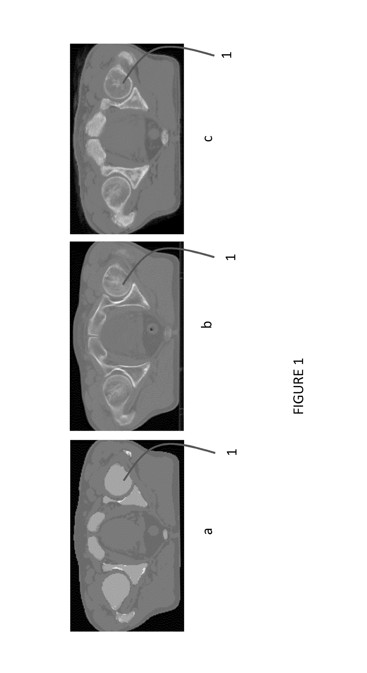
[0050]FIG. 1 shows a comparison between a pseudo CT image according to the prior art (a), a true CT image (b) and a pseudo CT image derived according to an embodiment of the invention (c). FIG. 1a is generated by average HU value assignment to water and fat (Dixon-reconstruction), air (body outline detection) and cortical and cancellous bone (segmentation+threshold). FIG. 1c is generated by interpolation of average CT values based on relative prevalence as proposed using bone segmentation. For the generation of FIG. 1c no body-outline detection was used. FIG. 1c shows a lot more detail in bone tisse 1 compared to FIG. 1a. Furthermore, FIG. 1c is visually more similar to FIG. 1b then FIG. 1a.

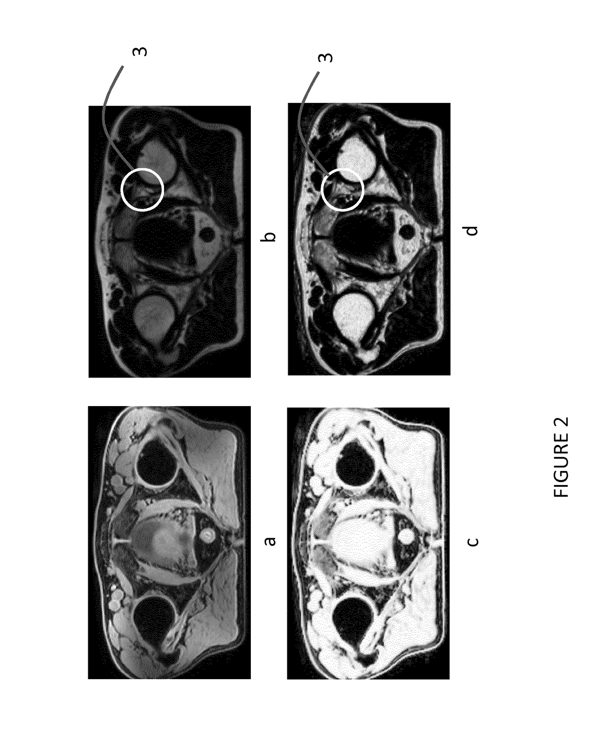
[0051]FIG. 2 shows intensity values in Dixon reconstructed ima...


PUM


Abstract

A pseudo CT Hounsfield Unit value for a volume element within a subject is estimated from a plurality of magnetic resonance images having different contrasts. The method includes determining a relative prevalence of a first tissue class and second tissue class within the volume element from a first magnetic resonance image and a second magnetic resonance image, respectively. Then a relative prevalence of a third tissue class is determined within the volume element based on substraction of a relative prevalence of the first and / or second tissue class from a total tissue prevalence. A reference Hounsfield Unit value is provided for the first, second and third tissue class. Finally, a pseudo Housfield value is estimated for the volume element by determining a weighted sum of the first, second and third reference Hounsfield unit value, with weight factors which are based on the determined relative prevalences of the first, second and third tissue class.

Description

CROSS REFERNCE TO RELATED APPLICATIONS[0001]This application is a U.S. national phase application of International Application No. PCT / EP2015 / 055539, filed on Mar. 17, 2015, which claims the benefit of EP Application Ser. No. 14163038.4 filed on Apr. 1, 2014 and is incorporated herein by reference.FIELD OF THE INVENTION[0002]The invention relates to magnetic resonace imaging, in particular to the creation of pseudo pseudo Housfield value for voxels.BACKGROUND OF THE INVENTION[0003]Creating estimated electron density maps from magnetic resonance (MR) images is a problem that occurs e.g. with integrated PET / MR systems, where an attenuation maps needs to be estimated, or with radiation therapy planning based on MR, where electron density maps are required for the treatment simulation. However, due to the physics of the image acquisition, MR intensities do not uniquely correspond to electron density, hence those maps cannot be derived from the MR image by a simple lookup operation, as i...

Claims


Application Information

Patent Timeline
Patent Type & Authority Patents(United States)
IPC IPC(8): G01R33/56G06T7/11G06T7/00G01R33/48A61B5/00A61B5/055
CPCG01R33/5608A61B5/0042A61B5/055G01R33/481G06T7/0012G06T7/11G01R33/4828G06T2207/30024G06T2207/10088G06T2207/30008G06T5/008G06T5/50
Inventor SCHADEWALDT, NICOLEHELLE, MICHAEL GUNTERSCHULZ, HEINRICHRENISCH, STEFFEN
Owner KONINKLJIJKE PHILIPS NV
Why Eureka
Try Eureka