
Method and system for enhanced imaging visualization of deep brain anatomy using infusion

a deep brain and imaging visualization technology, applied in the field of medical imaging, can solve the problems of small and indistinct, contemporary imaging systems and techniques are limited to demonstrate the anatomy, and inherent errors in targeting

Active Publication Date: 2018-12-25
UNIV OF VIRGINIA ALUMNI PATENTS FOUND
View PDF85 Cites 3 Cited by
  • Summary
  • Abstract
  • Description
  • Claims
  • Application Information

AI Technical Summary

Benefits of technology

The present invention provides technology and methods to visualize deep brain structures using infusates that highlight borders and internal patterns, enabling direct targeting without awake surgery. This reduces complications, enhances therapy, and serves to eliminate the need for awake surgery. The invention also includes a catheter device with associated lumens having diagnostic agent ports for delivering the diagnostic agent and imaging the brain site simultaneously. The diagnostic agent is able to highlight borders and internal patterns of the deep structures of the brain thereby enabling direct targeting.

Problems solved by technology

These structures can be very small and indistinct so that even contemporary imaging systems and techniques are limited to demonstrate the anatomy.
However, owing to the variation in the human brain from patient to patient, especially in patients with aging brains, or as a result of brain shift following opening of the skull, there are inherent errors in targeting.
Complications, adverse effects, and subtherapeutic results arise when the targeting of these structures is inadequate.
Many current methods and systems for MRI visualization of deep brain structures do not have the capability to visualize small, often indistinct regions that would otherwise greatly improve clinical outcomes with therapeutic interventions.
Many current methods and systems of stereotactic surgeries for interventions cannot reasonably rely on the accuracy of targeting deep brain anatomic structures like the subcortical nuclei.
Direct targeting is not possible owing to an inability to directly visualize the tissues of interest or final target.
This leads to errors that may require repeat surgery, increased risk of complications, and the need for awake surgery which can be frightening or intolerable to many patients.

Method used


Image

  • Method and system for enhanced imaging visualization of deep brain anatomy using infusion
  • Method and system for enhanced imaging visualization of deep brain anatomy using infusion
  • Method and system for enhanced imaging visualization of deep brain anatomy using infusion

Examples


example 1

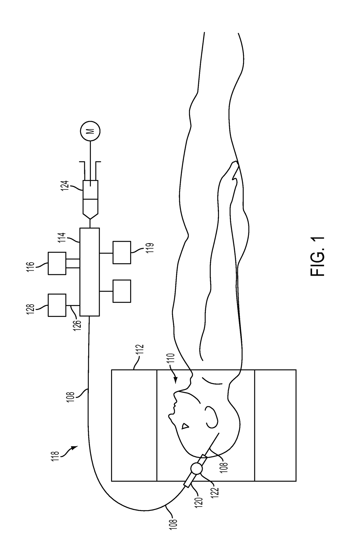
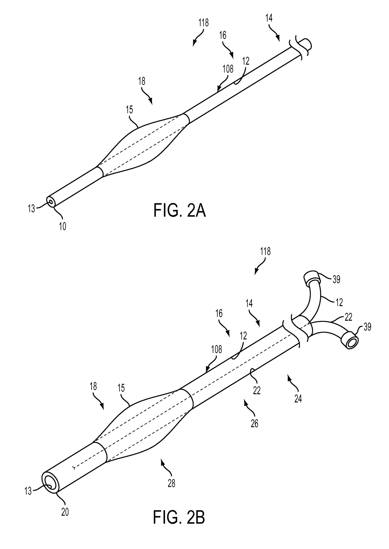
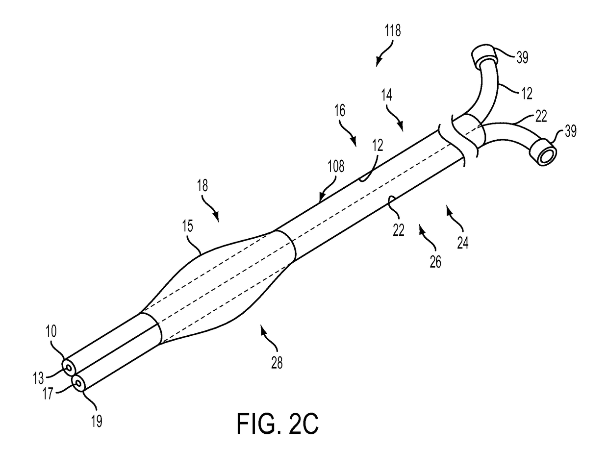
[0075]An aspect of an embodiment of the present invention provides, but not limited thereto, a catheter system for delivering a diagnostic agent to a site in the brain of a subject for imaging at least a portion of the brain site on a medical imaging system. The catheter system may comprise: a catheter device, the catheter device includes as a first lumen, the first lumen having a first lumen proximal region, a first lumen distal regional, and a first lumen longitudinal region there between; the first lumen configured to convey a diagnostic agent within the first lumen, and at least a portion of the first lumen having one or more ports configured to allow the conveyed diagnostic agent to exit from the first lumen to at least a portion of the brain site; and a portion of the catheter device having a cross-sectional area greater than portions of the catheter located proximally so as to define a seal within at least a portion of the brain site, wherein the seal is configured to prevent...

example 2

[0076]The system of example 1, further comprising:

[0077]a first lumen diagnostic agent, wherein the first lumen diagnostic agent comprises: autologous cerebrospinal fluid (CSF).

example 3

[0078]The system of example 1 (as well as subject matter of any combination of example 2), further comprising: a first lumen diagnostic agent, wherein the first lumen diagnostic agent comprises: artificial cerebrospinal fluid (CSF).


PUM


Abstract

Provided is a method and system for delivering a diagnostic agent to a site in the brain of a subject for imaging at least a portion of the brain site on a medical imaging system. The method and system includes a catheter device with associated lumens having diagnostic agent ports for delivering the diagnostic agent (e.g., infusate) through the lumens and advancing the diagnostic agent so as to exit out from the lumens to at least a portion of the brain site and while sealing a portion of the brain site thereby preventing the exited diagnostic agent from travelling proximally beyond the sealing location, and at the same time imaging at least a portion of the brain site during at least a portion of the sealing duration so that the brain site can be visualized on a medical imaging system. The diagnostic agent (infusate) is able to highlight borders and internal patterns of the deep structures of the brain thereby enabling direct targeting. Ultimately this leads to reduced complications, enhanced therapy, and the elimination of the need for awake surgery. The method and system provides the capability heretofore not possible to visualize the small, often indistinct regions, which will greatly improve clinical outcomes with therapeutic interventions.

Description

RELATED APPLICATIONS[0001]The present application is a national stage filing of International Application No. PCT / US2013 / 060404, filed Sep. 18, 2013, which claims priority under 35 U.S.C. § 119(e) from U.S. Provisional Application Ser. No. 61 / 703,191, filed Sep. 19, 2012, entitled “Enhanced Magnetic Resonance Visualization of Deep Brain Anatomy with Stereotactic Convection Infusion(s),” and U.S. Provisional Application Ser. No. 61 / 874,741, filed Sep. 6, 2013, entitled “Enhanced Magnetic Resonance Visualization of Deep Brain Anatomy with Stereotactic Convection Infusion(s);” the disclosure of which are hereby incorporated by reference herein in their entirety.TECHNICAL FIELD[0002]The present invention relates generally to the field of medical imaging. More specifically, the present invention also relates to a catheter device used to enhance deep brain magnetic resonance imaging using infusion.BACKGROUND[0003]Deep Brain anatomy has become the focus of many neurosurgical procedures whe...

Claims


Application Information

Patent Timeline
IPC IPC(8): A61B5/055A61N1/36A61B6/00A61B6/12A61M25/00A61M25/10A61M5/00A61B5/00A61B6/03G01R33/28A61M31/00A61M25/01
CPCA61M5/007A61B6/12A61B6/481A61B6/501A61M25/007A61M25/0026A61M25/10A61N1/36139A61B5/055G01R33/285A61B5/0042A61B6/032A61M25/0127A61M25/1002A61M31/005A61M2025/0037A61M2025/0042A61M2025/105A61M2025/1052A61M2025/1093
Inventor ELIAS, WILLIAM J.BOND, AARON E.GILLIES, GEORGE T.
Owner UNIV OF VIRGINIA ALUMNI PATENTS FOUND
Learn More