
Computer-implemented system and method for providing a personal mobile device-triggered medical intervention

Active Publication Date: 2019-01-01
BARDY DIAGNOSTICS
View PDF444 Cites 30 Cited by
  • Summary
  • Abstract
  • Description
  • Claims
  • Application Information

AI Technical Summary

Benefits of technology

The patent describes a wearable monitor for monitoring physiological data, including cardiac activity, in real-time or over time. The monitor has a unique shape that allows for comfortable and extended wear on the patient's chest, without causing discomfort or irritation to the breasts. It can be used with various wearable sensors and mobile devices, such as smartphones, and can also be equipped with a wireless transceiver for communication with external systems or further devices. The monitor recorder can authenticate the data recorded, and the monitor itself has features to prevent data corruption or unauthorized access. The technical effects of this invention include improved accuracy and diagnosis of arrhythmias, as well as increased comfort for female patients.

Problems solved by technology

ECGs are limited to recording those heart-related aspects present at the time of recording.
Thus, an ECG only provides a partial picture and can be insufficient for complete patient diagnosis of many cardiac disorders.
Although a 30-day observation period is considered the “gold standard” of ECG monitoring, achieving 30-day coverage has proven unworkable because conventional ambulatory ECG monitoring systems are arduous to employ, cumbersome to wear, and excessively costly.
For instance, the long-term wear of ECG electrodes is complicated by skin irritation and the inability of conventional ECG electrodes to maintain continual skin contact after a day or two.
These factors adversely affect electrode adhesion and the quality of cardiac signal recordings.
Moreover, physical movement and clothing impart compressional, tensile, and torsional forces on electrode contact points decreasing signal quality, especially over long recording times. In addition, an inflexibly fastened ECG electrode is prone to dislodgement that often occurs unbeknownst to the patient, making the ECG recordings worthless.
Further, some patients may have skin conditions, such as itching and irritation, aggravated by the wearing of most ECG electrodes.
Such replacement or slight alteration in electrode location can interfere with the goal of recording the ECG signal for long periods of time.
Finally, such recording devices are often ineffective at recording atrial electrical activity, which is critical in the accurate diagnosis of heart rhythm disorders, because of the use of traditional ECG recording electronics or due to the location of the monitoring electrodes far from the origin of the atrial signal, for instance, the P-wave.
A “looping” Holter (or event) monitor can operate for a longer period of time by overwriting older ECG tracings, thence “recycling” storage in favor of extended operation, yet at the risk of losing crucial event data.
Holter monitors remain cumbersome, expensive and typically for prescriptive use only, which limits their usability.
Further, the skill required to properly place the ECG leads on the patient's chest hinders or precludes a patient from replacing or removing the electrodes.
During monitoring, the battery is continually depleted and can potentially limit overall monitoring duration.
These patches have a further limitation because of a small inter-electrode distance coupled to its designed location of application, high on the left chest.
The electrical design of the ZIO patch and its location make recording high quality atrial signals (P-waves) difficult, as the location is relatively far from the origin of these low amplitude signals.
Furthermore, this patch is problematical for woman by being placed in a location that may limit signal quality, especially in woman with large breasts or bosoms.
Both ZIO devices represent compromises between length of wear and quality of ECG monitoring, especially with respect to ease of long term use, female-friendly fit, and quality of atrial (P-wave) signals.
An algorithm implemented as an app executed by the smartphone analyzed recorded signals to accurately distinguish pulse recordings during atrial fibrillation from sinus rhythm, although such a smartphone-based approach provides non-continuous observation and would be impracticable for long term physiological monitoring.
Further, the smartphone-implemented app does not provide continuous recordings, including the provision of pre-event and post-event context, critical for an accurate medical diagnosis that might trigger a meaningful and serious medical intervention.
In addition, a physician would be loath to undertake a surgical or serious drug intervention without confirmatory evidence that the wearer in question was indeed the subject of the presumed rhythm abnormality.
Furthermore, as explained supra with respect to the McManus reference, none of these devices provides the context of the arrhythmia, as well as the medico-legal confirmation that would otherwise allow for a genuine medical intervention.
The wearer must take extra steps to route recorded data to a health care provider; thus, with rare exception, the data is not time-correlated to physician-supervised monitoring nor validated.
Thus, such data that is available today is not actionable in a medically therapeutic relevant way.
To make such data actionable, one must have recorded data that allows a specific rhythm diagnosis, and a vague recording hinting that something may be wrong with the heart will not suffice.

Method used


Image

  • Computer-implemented system and method for providing a personal mobile device-triggered medical intervention
  • Computer-implemented system and method for providing a personal mobile device-triggered medical intervention
  • Computer-implemented system and method for providing a personal mobile device-triggered medical intervention

Examples


Embodiment Construction

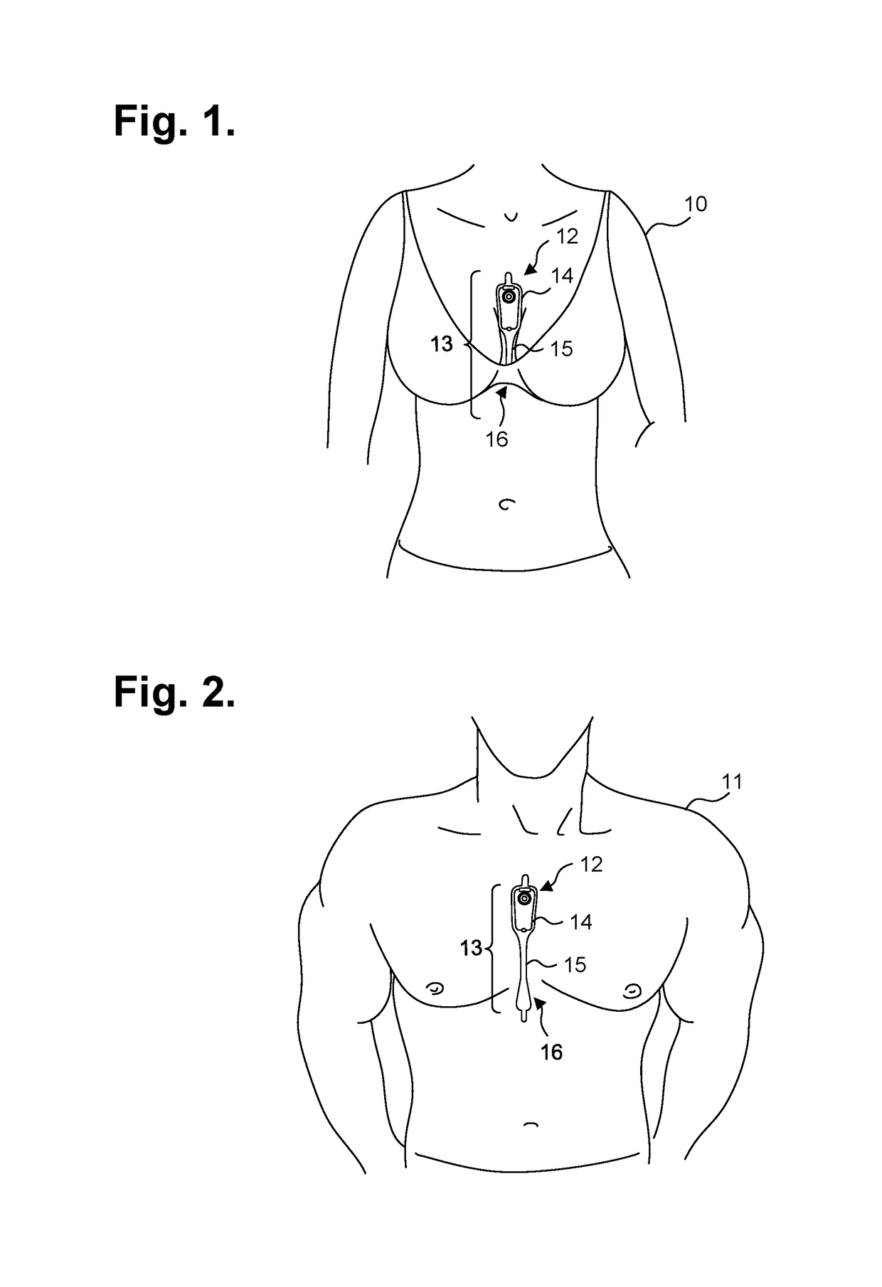
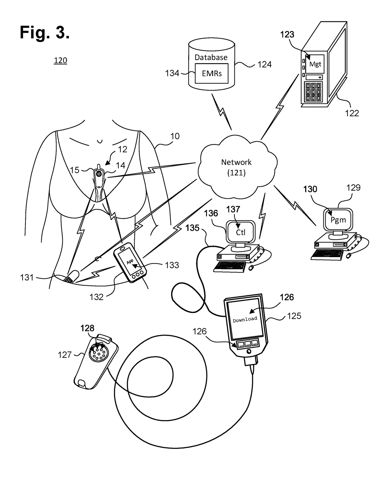
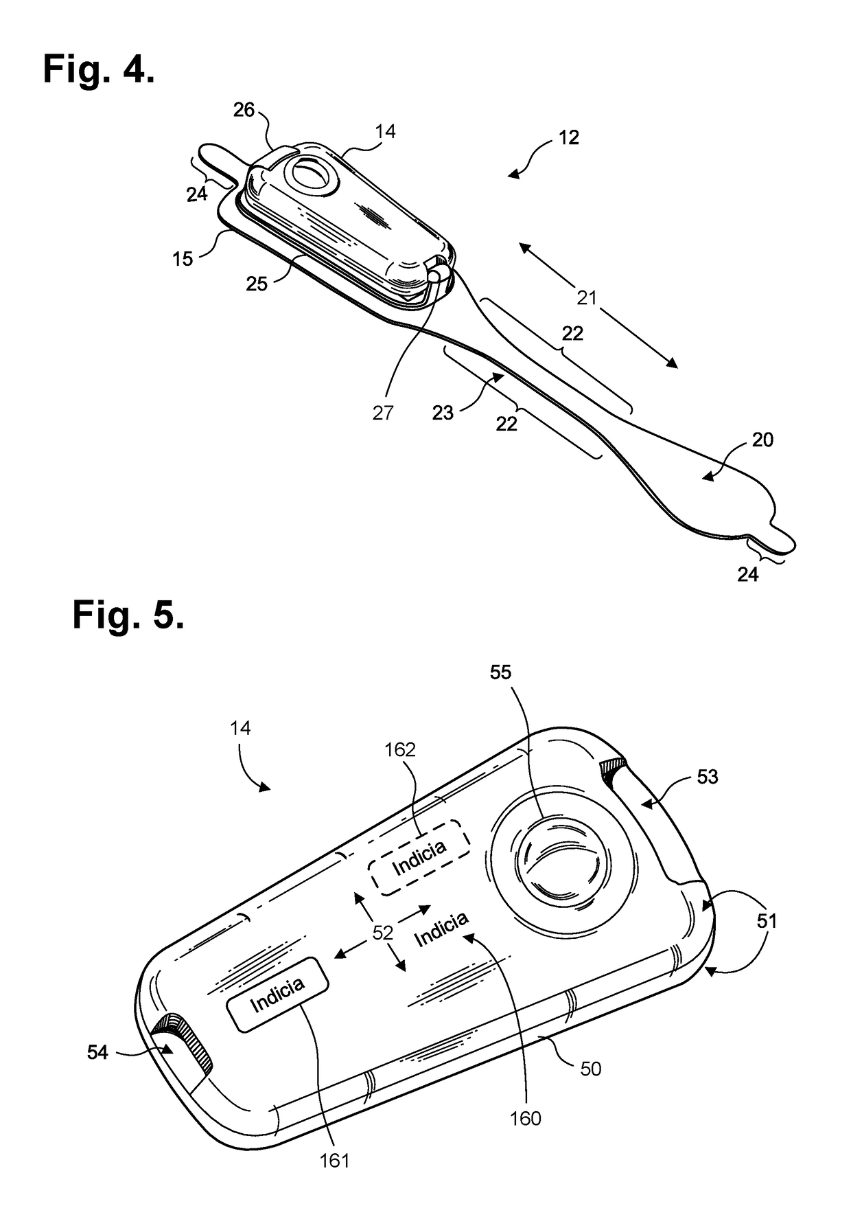
[0039]Physiological monitoring can be provided through a wearable monitor that includes two components, a flexible extended wear electrode patch and a removable reusable monitor recorder. FIGS. 1 and 2 are diagrams showing, by way of examples, an extended wear electrocardiography and physiological sensor monitor 12, including a monitor recorder 14 in accordance with one embodiment, respectively fitted to the sternal region of a female patient 10 and a male patient 11. The wearable monitor 12 sits centrally (in the midline) on the patient's chest along the sternum 13 oriented top-to-bottom with the monitor recorder 14 preferably situated towards the patient's head. In a further embodiment, the orientation of the wearable monitor 12 can be corrected post-monitoring, as further described infra. The electrode patch 15 is shaped to fit comfortably and conformal to the contours of the patient's chest approximately centered on the sternal midline 16 (or immediately to either side of the st...


PUM


Abstract

Physiological monitoring can be provided through a wearable monitor that includes two components, a flexible extended wear electrode patch and a removable reusable monitor recorder. The wearable monitor sits centrally on the patient's chest along the sternum oriented top-to-bottom. The placement of the wearable monitor in a location at the sternal midline (or immediately to either side of the sternum) benefits extended wear by removing the requirement that ECG electrodes be continually placed in the same spots on the skin throughout the monitoring period. The wearable monitor can interoperate wirelessly with other physiology and activity sensors and mobile devices. An application executed by the sensor or device can trigger the dispatch of a wearable monitor to the patient upon detecting potentially medically-significant events. The dispatched wearable monitor would then be capable of providing precise medically-actionable data, not merely an alert that some abnormality may be present. The patient can then use the sensor or device to physically record the placement and use of the medically-actionable wearable monitor, thereby facilitating the authentication of the data recorded.

Description

CROSS-REFERENCE TO RELATED APPLICATION[0001]This non-provisional patent application is a continuation-in-part of U.S. Pat. No. 9,433,367, issued Sep. 6, 2016; which is a continuation-in-part of U.S. Pat. No. 9,545,204, issued Jan. 17, 2017, and a continuation-in-part of U.S. Pat. No. 9,730,593, issued Aug. 15, 2017; and which further claims priority under 35 U.S.C. § 119(e) to U.S. Provisional Patent application, Ser. No. 61 / 882,403, filed Sep. 25, 2013, the disclosures of which are incorporated by reference.FIELD[0002]This application relates in general to electrocardiographic monitoring and, in particular, to a computer-implemented system and method for providing a personal mobile device-triggered medical intervention.BACKGROUND[0003]An electrocardiogram (ECG) records electrical potentials in the heart using a standardized set format 12-lead configuration to record cardiac electrical signals from well-established chest locations. Electrodes are placed on the skin over the anterior...

Claims


Application Information

Patent Timeline
IPC IPC(8): A61B5/02A61B5/024A61B5/0205A61B5/00G16H40/67G16Z99/00
CPCA61B5/0022A61B5/024G16H40/63A61B5/7282G06F19/00A61B5/0205G16H40/67G16Z99/00
Inventor BARDY, GUST H.FELIX, JASONBISHAY, JON MIKALSON
Owner BARDY DIAGNOSTICS
Learn More