Eureka AIR delivers breakthrough ideas for toughest innovation challenges, trusted by R&D personnel around the world.

Medial axis extraction for complex 3D objects

a technology of complex 3d objects and axes, applied in the field of medial axis extraction for complex 3d objects, can solve the problems of missing axis, missing skeleton, and missing skeleton,

Active Publication Date: 2019-10-08
UNIVERSITY OF MANITOBA
View PDF9 Cites 4 Cited by
  • Summary
  • Abstract
  • Description
  • Claims
  • Application Information

AI Technical Summary

Problems solved by technology

However, there are several drawbacks to this approach.
However, the main drawback is that the resulting medial axis may not be one-pixel thin, or that holes / discontinuities can sometimes appear in the resulting skeleton.
The primary drawback of this approach is that the extracted skeleton may contain additional / redundant information due to the presence of noise; however, this can be removed using various pruning methods [27][28][29].
Therefore, these edges are not considered to be spurious and are not eliminated from the final accurate skeleton.
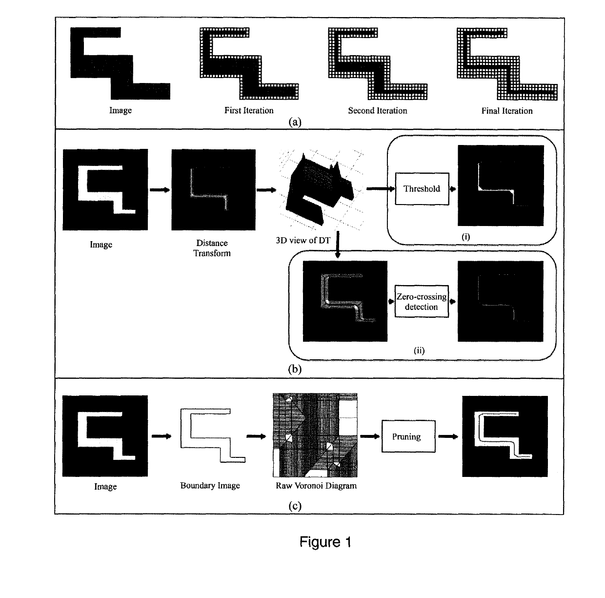
While the forgoing process is useful for deriving the medial axis for a 2D image, it is not directly applicable to determine the medial axis for a 3D object.

Method used


Image

  • Medial axis extraction for complex 3D objects
  • Medial axis extraction for complex 3D objects
  • Medial axis extraction for complex 3D objects

Examples


experiment 1

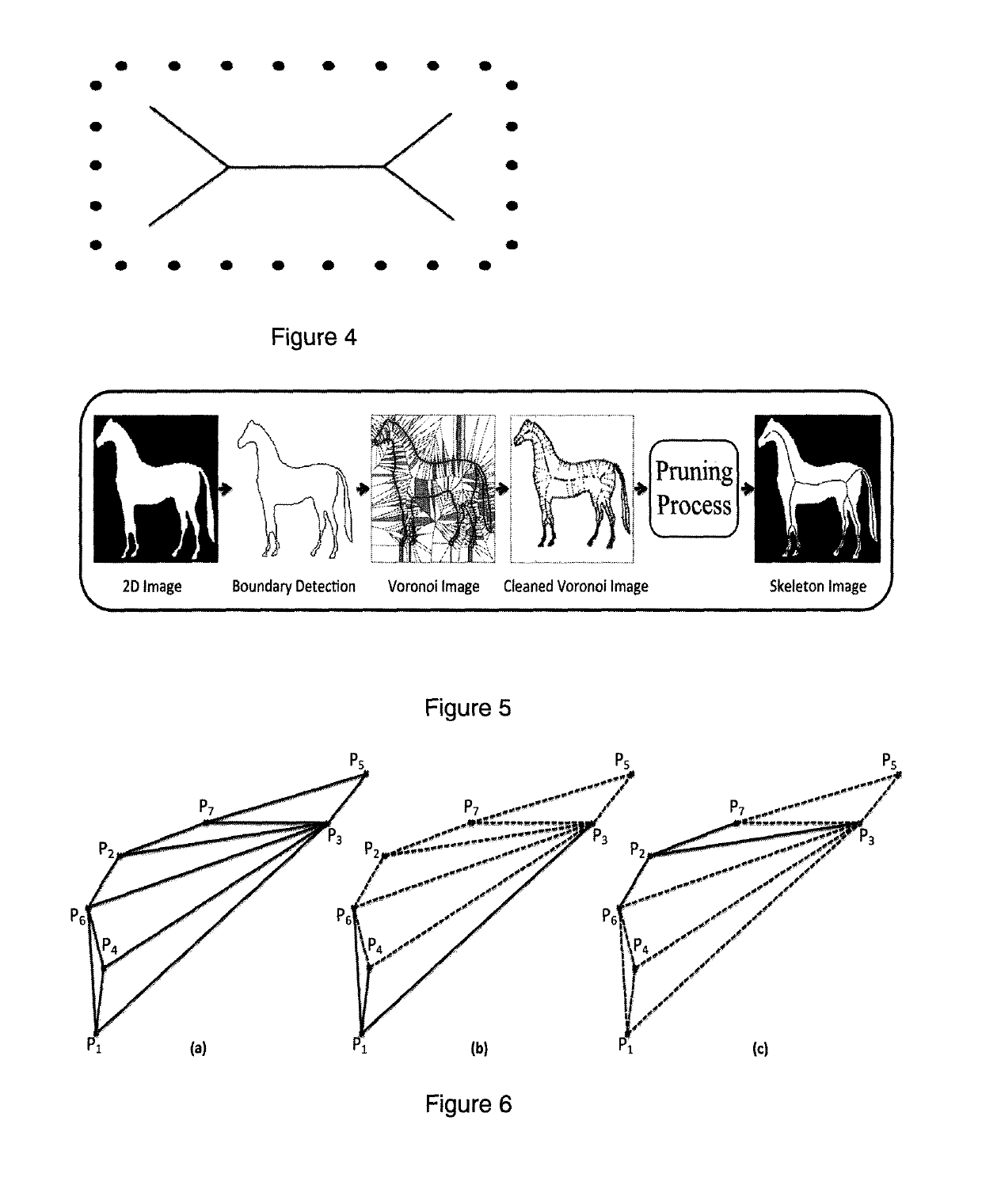
[0103]The control parameter T used in (3) controls the strictness of the pruning process by determining the number of branches that are retained in the final extracted skeleton. In this case, lower values of it (e.g., π / 3, π / 2, 2π / 3) will retain more branches in the skeleton, while using higher values of it (such as 2π) will remove more branches from the skeleton. FIG. 11 shows the effect of different values of π on the skeleton.

[0104]The stability of the Voronoi algorithm was then tested on a random assortment of 2D binary images taken from the benchmark MPEG-7 database [45]. For this experiment, different variations of control parameter were used in order to extract the skeleton of the respective image. FIG. 12 shows the results, which prove that the implemented 2D Voronoi algorithm accurately extracted the 2D skeletons for each image.

experiment 2

[0105]In this experiment, the complete Voronoi algorithm for 3D medial axis extraction was tested on a random assortment of smooth and continuous 3D binary images from the McGill 3D Shape Benchmark database [46]. For each 3D image, the algorithm produced four possible medial axes (FIG. 13), as discussed above in the methodology section. However, the most accurate among these (selected by visual examination) consistently demonstrated that the presently disclosed algorithm accurately extracted the connected medial axis and maintained the 3D topology of the original smoothed and continuous image (FIG. 14). With reference to FIG. 13, clearly The medial axis formed by the YZ-common intersection (b) is the only skeleton that is continuous, one voxel thin, and preserves the topology of the input image. Therefore, this is can be easily (visually) identified by the user and saved as the correct medial axis.

experiment 3

[0106]After confirming that the 3D medial axis extraction algorithm consistently performed well on smooth and continuous 3D images, it was further desirable to identify whether it was robust enough for potential 3D medical imaging applications. Binary masks from medical images can be more complex than the previous benchmarking images due to lower resolution and less smooth, more irregular shapes. As a result, extracting medial axes for these kinds of images is a more complicated task.

[0107]In this experiment, the 3D medial axis extraction algorithm was subjected to a ‘stress test’ by evaluating its performance on human brain white matter atlases that were derived from diffusion tensor imaging data. Specifically, a random assortment of fiber tracts were chosen from the University of Manitoba & Johns Hopkins University Functionally-Defined White Matter Atlas [47] to test the algorithm. As shown in FIG. 15, the algorithm worked surprisingly well for most of the fiber tracts. Among the ...


PUM


Abstract

A novel methodology for computing the medial axis / skeleton of a discrete binary object using a ‘divide and conquer’ algorithm, in which any 3D object is first sliced into a series of 2D images in X, Y and Z directions. Then, a geometric (Voronoi) algorithm is applied on each 2D image to extract the respective medial axis. This information is then combined to reconstruct the medial axis of the original 3D object using an intersection technique. An optional 3D interpolation step to achieve continuous connected skeletons uses Delaunay triangles and a spherical search to establish the nearest neighboring points in 3D space to interpolate between. Test results show that the proposed 3D Voronoi and optional interpolation algorithms are able to accurately and efficiently extract medial axes for complex 3D objects as well. Finally, an axis-smoothing algorithm using the same Delaunay triangle and spherical test is operable to remove unwanted noise from the extracted medial axis.

Description

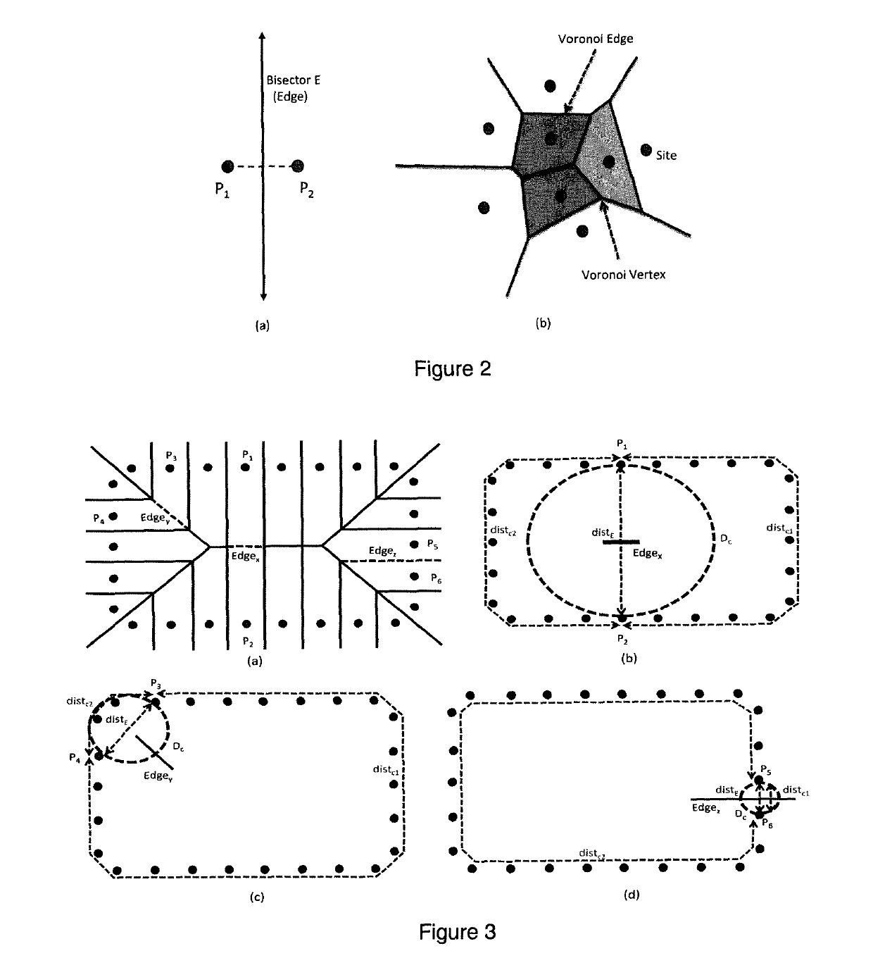
CROSS-REFERENCE TO RELATED APPLICATIONS[0001]This application claims benefit under 35 U.S.C. 119(e) of U.S. Provisional Application No. 62 / 435,311, filed Dec. 16, 2016, the entirety of which is incorporated herein by reference.BACKGROUND[0002]The medial axis (or “topological skeleton”) of an object, first described by Blum [1], can be defined as the set of points within the object that are equidistant from two or more closest points along the object's boundary. An accurate skeleton is a highly efficient way to represent the topology of 2D or 3D objects in a compact way [2][3] and has been widely used in various fields such as image processing and analysis, video graphics, character reconstruction, recognition and matching [4][5].[0003]Extraction of the medial axis or medial axis transform (MAT) for 2D / 3D object has typically fallen in one of the following three categories [6]: 1) boundary removal, 2) distance map suppression, or 3) geometric methods.[0004]Boundary removal, also know...

Claims


Application Information

Patent Timeline
Patent Type & Authority Patents(United States)
IPC IPC(8): G06T15/00G06T7/155G06T17/20G06T19/00G06T7/12
CPCG06T7/12G06T19/00G06T17/20G06T7/155G06T2219/008G06T2207/20044G06T2207/20041
Inventor YOUNAS, SOHAILFIGLEY, CHASE R.
Owner UNIVERSITY OF MANITOBA
Why Eureka
Eureka Blog
Learn More