System and method for tissue contact detection and for auto-exposure and illumination control

a tissue contact and auto-exposure technology, applied in the field of endoscopic imaging, can solve problems such as the quality of images provided, and achieve the effects of reducing the target brightness, and reducing the number of saturated pixels

Active Publication Date: 2019-12-24
INTUITIVE SURGICAL OPERATIONS INC
View PDF36 Cites 11 Cited by
  • Summary
  • Abstract
  • Description
  • Claims
  • Application Information

AI Technical Summary

Benefits of technology

[0019]The second control loop increases the output optical power and decreases the camera exposure time if the camera exposure time is larger than a first exposure threshold. The second control loop decreases the output optical power and increases the camera exposure time if the camera exposure time is smaller than a second exposure threshold. The second control loop leaves the output optical power and the camera exposure time unchanged if the camera exposure time is between the first exposure threshold and the second exposure threshold.
[0022]In still another aspect, the camera control unit includes an auto-exposure module. The auto-exposure module is coupled to the statistics module to receive the brightness histogram for a frame in the video stream. Also, the auto-exposure module is configured to determine an average brightness using the information in the histogram. The auto-exposure module is also configured to adjust one or both of a video pipeline gain and the camera exposure time based on a relation of the average brightness to the target brightness. The auto-exposure module also reduces the target brightness when a number of saturated pixels in a captured scene is larger than a saturated pixel threshold. In addition, the auto-exposure module is configured to command the illumination controller to change a first illuminator output to a second illuminator output and to command the camera to adjust the camera exposure to compensate for the change in illuminator output.
[0023]A method of operating a teleoperated surgical system includes adjusting one or both of a video pipeline gain and a camera exposure time using an average brightness of a captured scene and a target brightness for a displayed scene. The method also configures output optical power of an illuminator and exposure time of a camera using the camera exposure time. The method reduces a number of saturated pixels in a second captured frame by reducing the target brightness.

Problems solved by technology

However, the illumination of a surgical site and the exposure time of a camera used during capture of an image of a surgical site are some of the factors affecting the quality of the images provided to the surgeon.
However, a potential problem with a high intensity light source has been recognized and a solution provided in U.S. Pat. No. 6,511,422 B1.

Method used


Image

  • System and method for tissue contact detection and for auto-exposure and illumination control
  • System and method for tissue contact detection and for auto-exposure and illumination control
  • System and method for tissue contact detection and for auto-exposure and illumination control

Examples


Embodiment Construction

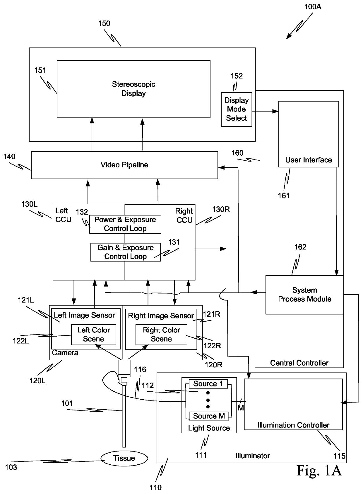
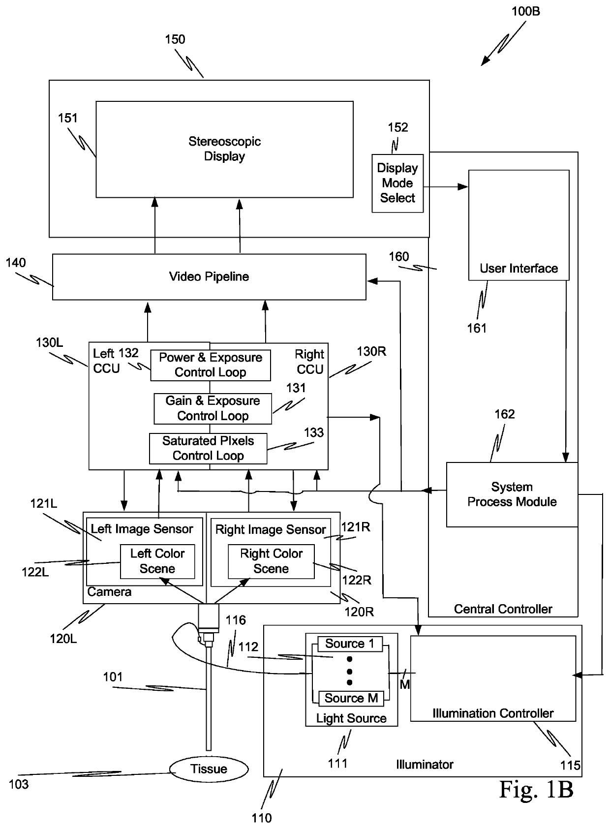
[0034]In one aspect, scenes captured with endoscope101 of teleoperated surgical systems 100A and 100B (FIGS. 1A and 1B) and displayed on stereoscopic display unit 151 maintain a consistent brightness even though the working distance between tissue 103 and the distal tip of endoscope 101 changes. Teleoperated surgical systems 100A and 100B also automatically detect when endoscope 101 contacts tissue and adjusts the output optical power of illuminator 110 so that tissue damage does not occur.

[0035]Illumination having an output optical power, e.g., visible light, from an illuminator 110 exits endoscope 101 at a distal tip of endoscope 101, sometimes referred as a tip. The illumination is reflected from tissue 103 and is captured as frame 122L that includes a left color scene captured by left image sensor 121L of left camera 120L and as a frame 122R that includes a right color scene captured by right image sensor 121R of right camera 120R. Each of left and right image sensors 121L and 1...


PUM


Abstract

Scenes captured with an endoscope (201) of a teleoperated surgical system (200) and displayed on a display unit (251) maintain a consistent brightness even though optical output power of an illuminator (210) changes, and working distance (204) between tissue (203) and the distal tip of the endoscope changes. Teleoperated surgical system (200) also automatically detects when endoscope (201) contacts tissue (203) and adjusts the output optical power of illuminator (210) so that tissue damage does not occur.

Description

RELATED APPLICATIONS[0001]This application is the U.S. national phase of International Application No. PCT / US2015 / 020892, filed Mar. 17, 2015, which designated the U.S. and claims priority to U.S. provisional patent application 61 / 954,336, filed on Mar. 17, 2014, and to U.S. provisional patent application 61 / 954,381, filed on Mar. 17, 2014, the contents of each of which are incorporated herein by reference.BACKGROUND[0002]Field of Invention[0003]Aspects of this invention are related to endoscopic imaging and are more particularly related to tissue contact detection, displayed scene brightness control, camera auto-exposure control, and illumination control in a teleoperated surgical system.[0004]Related Art[0005]The da Vinci® Surgical System, commercialized by Intuitive Surgical, Inc., Sunnyvale, Calif., is a minimally invasive teleoperated surgical system that offers patients many benefits, such as reduced trauma to the body, faster recovery and shorter hospital stay. One feature of...

Claims


Application Information

Patent Timeline
Patent Type & Authority Patents(United States)
IPC IPC(8): A61B1/00A61B34/30A61B1/045A61B90/30A61B1/06A61B90/00
CPCA61B34/30A61B1/045A61B1/00193A61B1/00006A61B1/06A61B1/00062A61B1/00165A61B1/00009A61B90/30A61B2090/08021A61B2090/065A61B2090/0801A61B2034/301A61B1/00055A61B1/0655A61B34/35
Inventor RICHMOND, GEOFFHOFFMAN, BRIAN D.DICARLO, JEFFREY M.
Owner INTUITIVE SURGICAL OPERATIONS INC
Why Eureka
Try Eureka