Dual vacuum device for medical fixture placement including for thoracoscopic left ventricular lead placement

a vacuum device and thoracoscopic technology, applied in the field of dual vacuum devices for medical fixture placement including thoracoscopic left ventricular lead placement, can solve the problems of inability to achieve the effect of guiding the feet, and difficulty in achieving endocardial techniqu

Active Publication Date: 2020-01-07
DATTA SUBHAJIT
View PDF6 Cites 1 Cited by
  • Summary
  • Abstract
  • Description
  • Claims
  • Application Information

AI Technical Summary

Benefits of technology

[0025]It is preferred that the elongated body sheath be provided with a handle portion extending laterally therefrom for ease of manual use, though it will be appreciated that the invention may be adapted for robotic use. The handle portion, such as a handle extending laterally from the elongated body, allows the operator to advance the device into the surgical site, and further articulate the inner and outer tubular vacuum guides allowing ultimately for the lead drive to insert the cardiac lead through operation of the lead placement device, as described herein.
[0039]The device may also additionally comprise an interferant release collar attached to the inner tubular lead guide distal end portion and disposed on the distal side of the inner guide suction foot portion, the interferant release collar being larger than the aperture. This allows more precise and oriented lead placement through advancement of the lead head portion distally from the inner tubular lead guide, while preventing accidental or premature retraction of the lead head portion, as described herein.
[0057]It will be appreciated that in another variation of the invention, that a flexible and resilient conduit portion may extend from the distal end of the hollow conduit guide and thereby also serve as a flexible attachment permitting the suction feet to be articulated while still being of sufficient strength to steer the feet.

Problems solved by technology

This total includes the cost of health care services, medications, and lost productivity.
A delay between the contraction of the right and left ventricles often occurs with heart failure, so the walls of the left ventricle are unable to contract synchronously.
The endocardial technique is technically challenging.
In some cases, this technique may not be successful due to the size, shape or location of the vein(s).
There are several complications and costs associated with conventional pacing that may occur during biventricular pacing, including: (a) localized / skin infection, (b) systemic infection secondary to infected pacing box or lead, (c) bleeding, (d) hematoma, (e) lead displacement, (f) equipment failure, i.e. fractured pacing wire, faulty pacing box and (g) pneumothorax.
Although with experience the procedure times reduce, even in the best hands implantation of the left ventricular lead may be time consuming, contributing to an increased infection risk.3 3 Alonso, C, et al.
These approaches required a larger incision and general anaesthetic, consequently carrying a significant morbidity and mortality.
Some of these procedures may require the use of multiple types of catheters and guidewires, adding cost to this procedure.
The requirement to position a lead in a branch of the coronary sinus and the techniques required to achieve this account for the additional complications and significant failure rate seen with biventricular pacing.
A suitable vein may not be present in the lateral or posterolateral position, prompting placement of left ventricular lead in another ineffective suboptimal location.
Trans-cardiac-venous placement of left ventricular lead occasionally will cause disturbing phrenic nerve stimulation, causing uncomfortable diaphragmatic twitching and necessitate relocation to suboptimal sites.

Method used


Image

  • Dual vacuum device for medical fixture placement including for thoracoscopic left ventricular lead placement
  • Dual vacuum device for medical fixture placement including for thoracoscopic left ventricular lead placement
  • Dual vacuum device for medical fixture placement including for thoracoscopic left ventricular lead placement

Examples


Embodiment Construction

[0104]In accordance with the foregoing summary, the following provides a detailed description of the preferred embodiment, which is presently considered to be the best mode thereof.

[0105]As used herein the distal end refers to the working end or patient end, while the proximal end refers to the operator end or actuator end from which the device of the present invention may be operated. The handle as shown in the described embodiment is on the side of the device referred to as the bottom side or the ventral aspect. The side opposite the bottom side is referred to as the top side or dorsal aspect. The right side is the side on the right hand when looking from the operator end, end-on. Conversely, the left side is the side on the left hand when looking from the operator end, end-on.

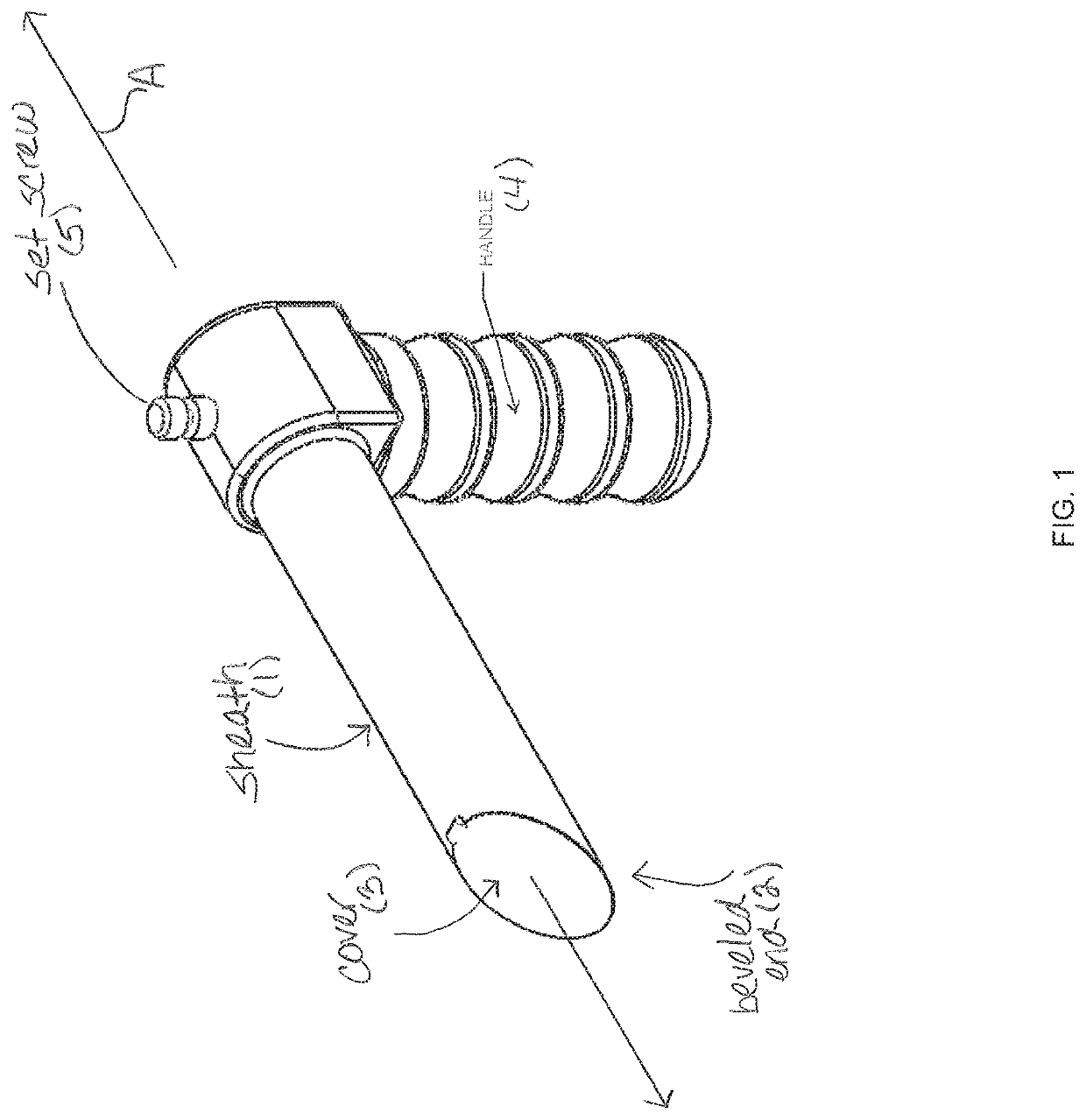
[0106]FIG. 1 is a first side lateral perspective view of an elongated sheath body 1 for a device in accordance with one embodiment of the present invention, having longitudinal axis A extending from the prox...


PUM


Abstract

The present invention includes devices and methods for lead, conduit or other medical fixture placement in tissues or organs. The device is configured to permit the placement foot, such as a suction foot, to articulate to a desired position with respect to the target tissue, while the lead, conduit or other medical fixture is releasably attached to the placement foot to permit it to be released from the placement foot after stabilization on the target tissue site. In a preferred embodiment, the invention features an articulating dual suction foot device, an inner lead conduit or guide and foot contained within an outer lead conduit or guide and foot, with the inner conduit or guide configured to extend from the outer conduit or guide, and to be further articulated once extended.

Description

RELATED APPLICATION DATA[0001]This application is a continuation of U.S. application Ser. No. 14 / 223,562, filed Mar. 24, 2014, now U.S. Pat. No. 9,511,219, issued Dec. 6, 2016, which is hereby incorporated in its entirety herein by reference.FIELD OF THE INVENTION[0002]The present invention relates to devices and techniques used in thoracoscopic lead placement, and in similar surgical techniques.BACKGROUND OF THE INVENTION[0003]According to a CDC statistical brief, nearly 5.8 million people in the United States have congestive heart failure. See Lloyd-Jones D, et al. Heart Disease and Stroke Statistics-2010 Update. A Report from The American Heart Association Statistics Committee And Stroke Statistics Subcommittee. Circulation. 2010; 121:E1-E170. About 670,000 people are diagnosed with it each year. Heart failure was a contributing cause of 282,754 deaths in 2006. In 2010, heart failure cost the United States an estimated $39.2 billion. This total includes the cost of health care se...

Claims


Application Information

Patent Timeline
Patent Type & Authority Patents(United States)
IPC IPC(8): A61N1/05A61N1/372A61B18/14A61B18/00
CPCA61B18/14A61N1/059A61B18/1492A61N1/0592A61N1/37205A61B2018/1475A61B2018/00357A61B2018/1412A61B2018/00291A61B2218/007A61B2018/00607A61B2018/00595A61B2018/00351A61N1/0587A61B2017/00243
Inventor DATTA, SUBHAJIT
Owner DATTA SUBHAJIT
Why Eureka
Try Eureka