Biomarker detection methods and systems and kits for practicing same

a biomarker and detection method technology, applied in the field of biomarker detection methods and systems for practicing same, can solve the problems of poor sensitivity and unsuitable high throughput multiplex analysis

Active Publication Date: 2020-01-07
THE BOARD OF TRUSTEES OF THE LELAND STANFORD JUNIOR UNIV
View PDF38 Cites 2 Cited by
  • Summary
  • Abstract
  • Description
  • Claims
  • Application Information

AI Technical Summary

Problems solved by technology
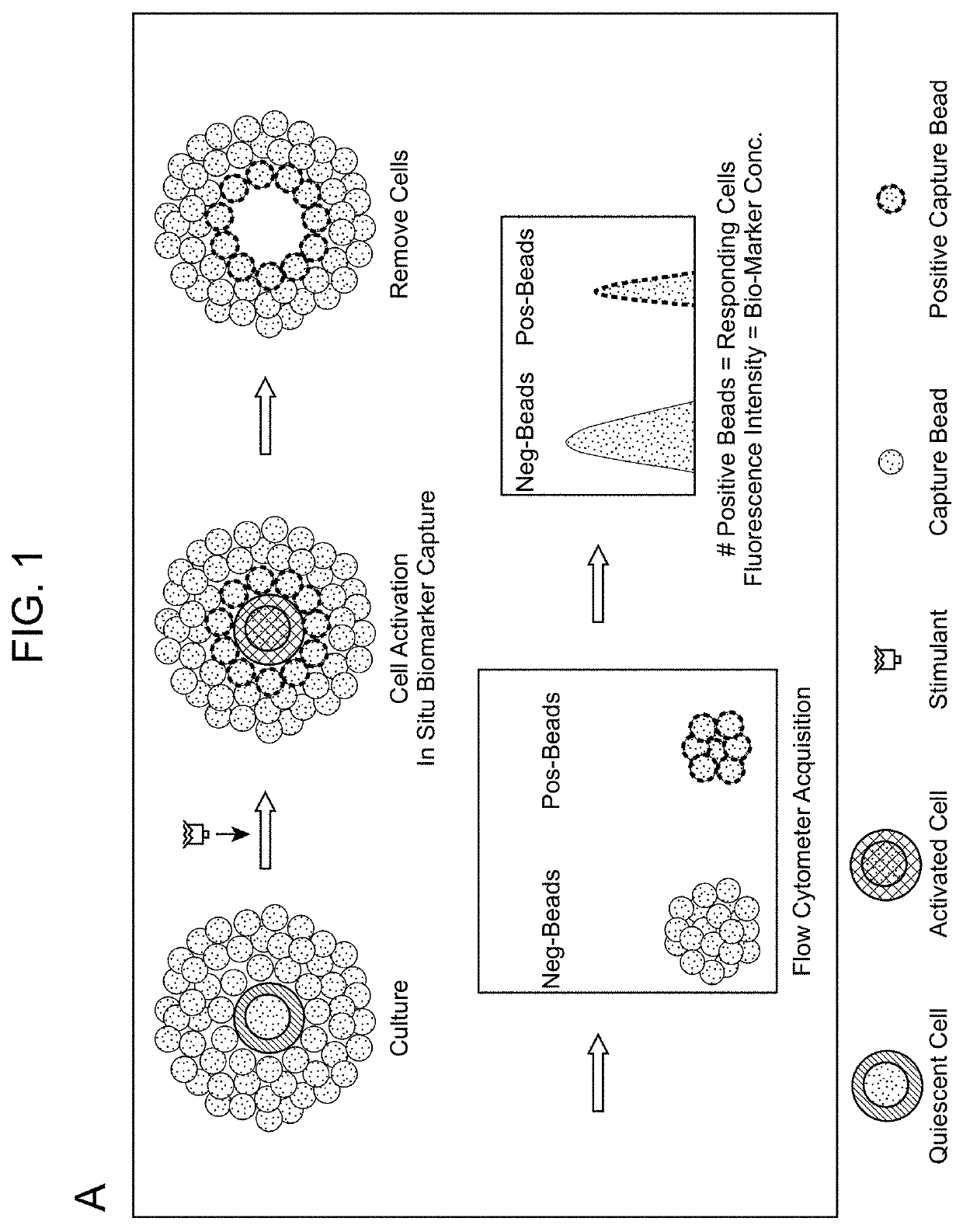
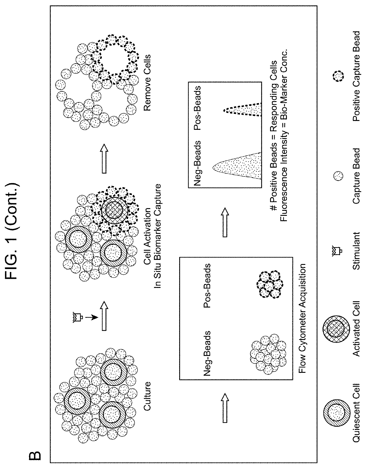
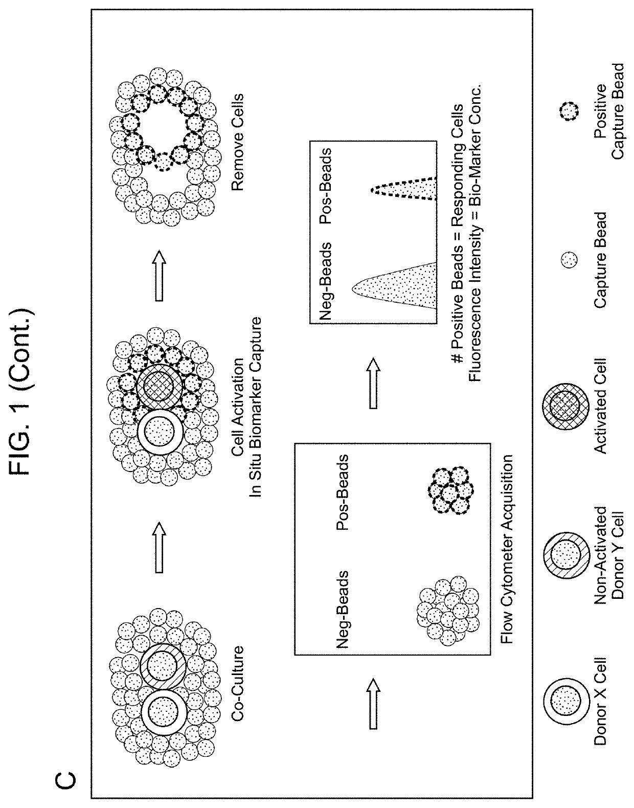
ELISA-based assays have long been the standard for quantitative analysis of cytokines and other biomarkers, but are not well suited for high throughput multiplex analyses and suffer from poor sensitivity due to dilution by diffusion in the supernatant.

Method used


Image

  • Biomarker detection methods and systems and kits for practicing same
  • Biomarker detection methods and systems and kits for practicing same
  • Biomarker detection methods and systems and kits for practicing same

Examples


example 1

of Phytohaemagglutinin (PHA) Induced Interferon Gamma (IFNγ) Spots by ELISPOT and FlowSpot

ELISPOT

[0123]One hundred thousand PBMC cells from C.T.L. (Cat# CTL-QC1, Shaker Heights, Ohio) were plated into a filter plate well pre-coated with IFNγ capture antibody and cultured for 16 h in the presence of PHA at various concentrations. The positive IFNγ spots were enumerated using a C.T.L. ImmunoSpot® Analyzer (FIG. 3 and FIG. 4).

FLOWSPOT

[0124]One hundred thousand of the same PBMC cells used in ELISPOT procedure were co-cultured with 7,000 IFNγ capture microparticles for 16 h in the presence of PHA at various concentrations. The spots and fluorescence intensity (FI) of FLOWSPOT were counted and measured by a FACSCanto II flow cytometer (BD Biosciences). Total number of positive IFNγ spots were calculated based on the previously described formulas. The FLOWSPOT was observed to be at least twenty times more sensitive than ELISPOT and able to detect very weak IFNγ responses induced by PHA (FI...

example 2

pecific IFNγ Spots Detection by FLOWSPOT

[0125]One hundred thousand HLA-typed antigen specific T cells from C.T.L. (Cat# CTL-QC1, Shaker Heights, Ohio) were co-cultured with 7,000 IFNγ capture microparticles for 16 h in the presence of the different peptides (10 nM) specified in FIG. 5. The IFNγ secretion induced by three peptides (#Cat. CTL-QC1; C.T.L.) were previously tested against the paired PMBCs (C.T.L. cells) by ELISPOT assay in C.T.L. and showed that HCMV pp65, Flu-Matrix, and influenza A were able to elicit strong, medium, and negative IFNγ responses on the PBMCs respectively. The same response pattern was also observed using FLOWSPOT but yielded a two-parameter result collected simultaneously in the same reaction well, i.e., the percentage of positive responding cells (% positive spots) and the relative IFNγ concentration (FI). As shown in FIG. 5, HCMV pp65 induced 19% IFNγ positive spots (1,330 spots) with a high FI (FI=25,286) and Flu-Matrix induced 20% positive spots (1,...

example 3

IFNγ / IL-2 Detection by FLOWSPOT

[0126]One hundred thousand C.T.L. cells were co-cultured with a mix of IFNγ and IL-2 capture microparticles (7,000 each with different fluorescent ID codes) for 16 h in the presence of PHA at various concentrations. FIG. 6 shows the dose-dependent responses of IFNγ and IL-2 to polyclonal T cell stimulus PHA.


PUM

PropertyMeasurementUnit
temperatureaaaaaaaaaa
pHaaaaaaaaaa

Abstract

Aspects of the present disclosure include methods that include co-culturing a cell and a microparticle that includes a capture ligand, in a culture medium under conditions in which a biomarker produced by the cell is bound by the capture ligand. Such methods may further include detecting (e.g., by flow or mass cytometry) complexes that include the microparticle, the capture ligand, the biomarker, and a detection reagent. The methods may further include determining the proportion or number of cells among a heterogeneous cell population that produced the biomarker and / or the level of biomarker secreted by such cells. Compositions, systems and kits are also provided.

Description

CROSS-REFERENCE TO RELATED APPLICATIONS[0001]Pursuant to 35 U.S.C. § 119 (e), this application claims priority to the filing date of the U.S. Provisional Patent Application Ser. No. 62 / 253,549, filed Nov. 10, 2015, the disclosure of which is herein incorporated by reference.INTRODUCTION[0002]The measurement of soluble and secreted cytokines and other analytes in serum and plasma is becoming increasingly important in the study and management of many diseases. Immune cells can be stimulated by various means to produce specific analytes. Secreted analytes can be quantitatively measured by ELISA or Cytometric Bead Array (CBA). The frequencies of cytokine-producing cells can be measured by Enzyme-Linked Immunospot (ELISPOT) or by flow cytometric analysis of intracellular cytokines.[0003]The ELISPOT assay is a widely used method for monitoring cellular immune responses in humans and other animals, and has found clinical applications in the diagnosis of tuberculosis and the monitoring of g...

Claims


Application Information

Patent Timeline
Patent Type & Authority Patents(United States)
IPC IPC(8): G01N33/50C12N5/00G01N33/543
CPCC12N5/0075G01N33/54313C12N2531/00G01N2333/705G01N33/50C12Q1/25G01N33/5047
Inventor TYAN, DOLLY B.CHEN, GE
Owner THE BOARD OF TRUSTEES OF THE LELAND STANFORD JUNIOR UNIV
Why Eureka
Try Eureka