

Compositions and methods for the treatment of neuromyelitis optica
a technology of optic nerve and composition, applied in the field of immunology, neurology and pathology, can solve the problems of limited treatment of symptoms, and achieve the effects of reducing one or more of retinal ganglion cell death, preventing or reducing the exacerbation of neuromyelitis optica (nmo) spectrum disease, and positive nmo-igg (aqp4) serology
- Summary
- Abstract
- Description
- Claims
- Application Information
AI Technical Summary
Benefits of technology
Problems solved by technology
Method used
Image

Examples
example 1
and Methods
[0172]DNA Constructs, Cell Culture and Transfections.
[0173]DNA constructs encoding full-length human AQP4 (M1 and M23 isoforms) were generated by PCR-amplification using whole brain cDNA as template. For some studies a Myc epitope (NH2-EQKLISEEDL-COOH; SEQ ID NO:13) was inserted in the second extracellular loop by PCR-amplification using the non-tagged constructs as template. Mutants of M1 and M23 were generated by PCR-amplification using either tagged or non-tagged templates. All PCR fragments were ligated into mammalian expression vector pcDNA3.1 and fully sequenced.
[0174]U87MG cell cultures (ATCC HTB-14) were maintained at 37° C. in 5% CO2 / 95% air in EMEM medium containing 10% fetal bovine serum, 100 U / mL penicillin and 100 μg / mL streptomycin. Cells were grown on glass coverslips and transfected with DNA in antibiotic-free medium using Lipofectamine 2000 (Invitrogen, Carlsbad, Calif.) according to the manufacturer's protocol. Stable AQP4-expressing clones were selected...
example 2
[0185]Approach for Quantitative Analysis of NMO-IgG Binding to AQP4.
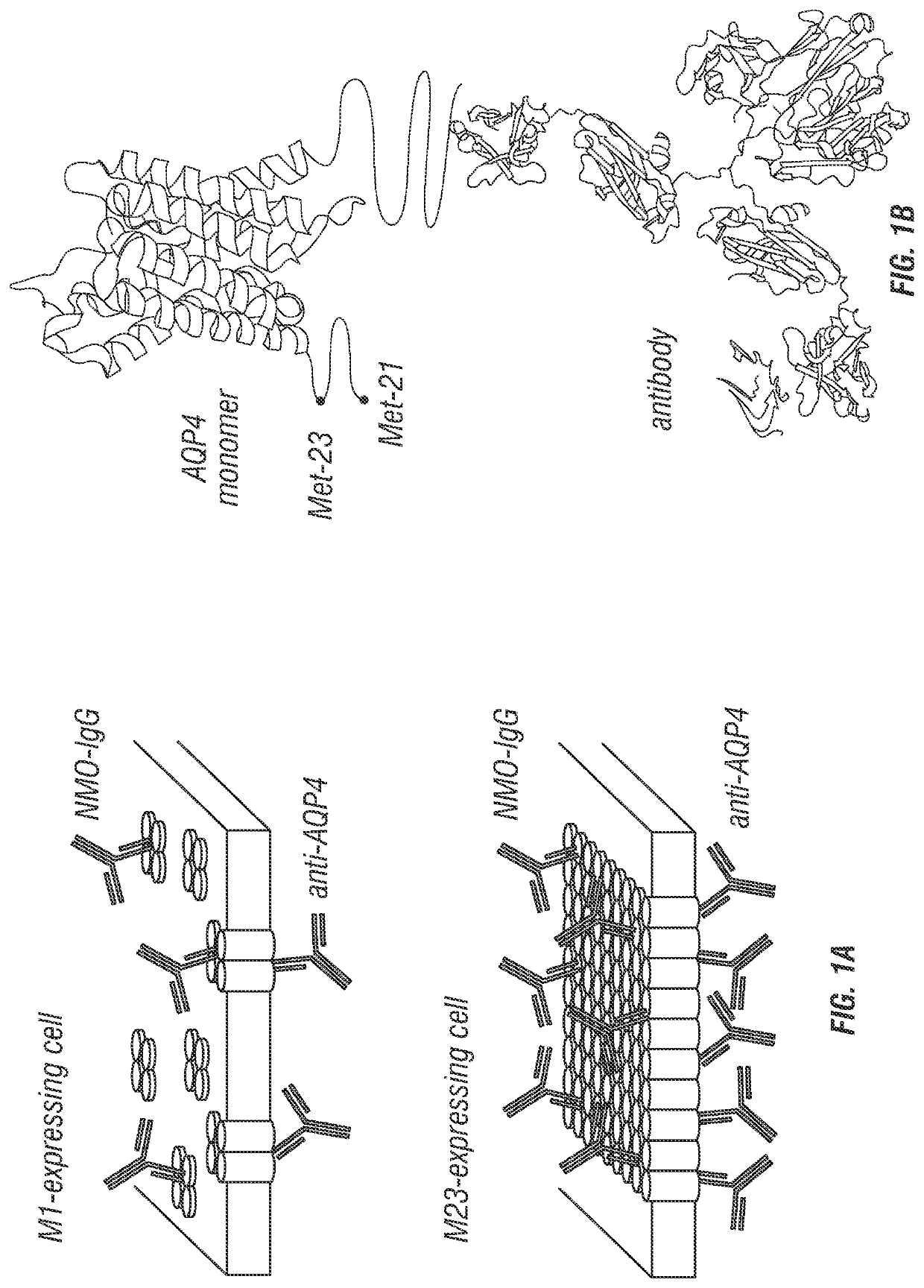
[0186]FIG. 1 diagrams the approach used for quantitative analysis of NMO-IgG binding to AQP4. Cells expressing specified AQP4 isoform(s) at their plasma membrane were incubated with NMO-IgG (NMO patient serum or recombinant monoclonal antibody), fixed, permeabilized, and then incubated with anti-AQP4 antibody (FIG. 1A). The anti-AQP4 antibody recognizes the AQP4 C-terminus, which is common to all AQP4 isoforms (FIG. 1B). Fluorescent secondary antibodies were used to detect NMO-IgG by green fluorescence and AQP4 by red fluorescence. NMO-IgG binding to AQP4 was quantified by green-to-red (G / R) fluorescence ratio. Binding affinity and stoichometry was determined by titration with increasing NMO-IgG concentration. In contrast to measurements done at a single NMO-IgG concentration, measurement of full, concentration-dependent binding provides a quantitative, unbiased description of NMO-IgG binding to AQP4.
[0187]A cell li...
example 3
n
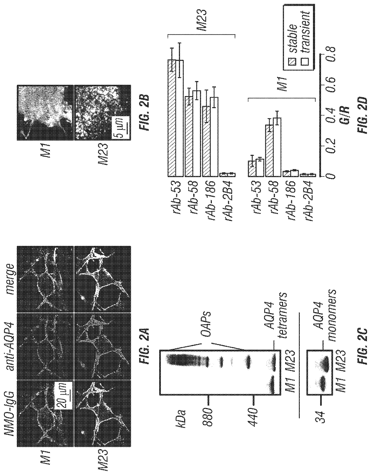
[0199]Here, the inventors used fluorescence ratio imaging to quantify the binding of NMO-IgG to AQP4, and to determine the role of AQP4 isoforms and OAPs in NMO-IgG binding. This live-cell system was developed out of a need for a robust method to characterize monoclonal NMO-IgGs and polyclonal NMO patient sera. The inventors found that U87MG cells were suitable for quantitative binding measurements because they efficiently expressed AQP4 at the plasma membrane after stable or transient transfection, with little or no intracellular AQP4 expression. The excellent membrane expression of AQP4 in U87MG cells is likely due to their glial origin, and hence their expression of the same trafficking machinery as that in native human glial cells. Immunoblot analysis of the stably transfected clones used in this study shows exclusive expression of individual AQP4 isoforms, with no detectable M23 in the M1 cell line (FIG. 2C). U87MG cells grow rapidly and adhere well to culture supports, making...
PUM
| Property | Measurement | Unit |
|---|---|---|
| concentration | aaaaa | aaaaa |
| temperatures | aaaaa | aaaaa |
| temperatures | aaaaa | aaaaa |
Abstract
Description
Claims
Application Information