Method and system for magnetic resonance imaging using sampled portions of K-space data

a magnetic resonance imaging and k-space data technology, applied in image data processing, diagnostics, applications, etc., can solve the problems of inability to readily match x-ray images and mr images, and achieve the effect of shortening the overall process time or acquisition time, reducing system wear and tear, and simplifying simultaneous acquisition of respective data

Active Publication Date: 2020-06-30
SIEMENS HEALTHCARE GMBH
View PDF3 Cites 0 Cited by
  • Summary
  • Abstract
  • Description
  • Claims
  • Application Information

AI Technical Summary

Benefits of technology

[0018]Since the MR data set acquired using the presently described method only and exactly contains the subset of the complete k-space data of the object needed for or corresponding to the final fan- or cone-beam geometry MR image, there is no need for a complex data selection step. Also, by sampling only the limited portion of the k-space needed for the final MR image, acquisition of the MR data set as well as the necessary processing steps, such as the inverse Fourier transformation, may be performed significantly faster as compared to traditional methods, where the complete k-space of the object is sampled and therefore a much larger MR data set is to be processed. The present embodiments therefore enable substantially real-time acquisition, processing, and displaying of MR images that are directly comparable and compatible to standard X-ray images. Since the final MR image and an X-ray image reconstructed from the acquired X-ray image data may have or correspond to the same beam or projection geometry, this obviates the need for a complex and time-consuming registration step that is necessary in conventional methods for correlating the X-ray and MR images. Not only do the present embodiments enable a direct superposition of the X-ray and MR images, but the present embodiments do so with reduced data acquisition effort, reduced computational effort, and reduced processing time.
[0037]In a development of the system, a magnet of the MR device, which creates a magnetic field for the magnetic resonance imaging process, is movably mounted, so that the magnet may be tilted with respect to the X-ray device or X-ray imaging device. This may improve a range of angulations accessible for imaging or data acquisition to the X-ray device and / or the MR device. If, for example, a head of a patient is to be examined, but the X-ray device may not be set to a cranial-caudal angulation due to limitations in the range of motion of the system or the X-ray device, the magnet of the MR device may be tilted to capture or measure the corresponding MR data used for creating or generating the final MR image with a perspective corresponding to the desired cranial-caudal angulation. Additionally or alternatively, the magnet of the MR device may be tilted out of the way of the X-ray device to allow for a greater range of motion of the X-ray device. For example, the magnet of the MR device may be tiltable by at least 10° in each direction or in two opposing directions.

Problems solved by technology

It is, however, problematical that known methods result in MR images based on a parallel beam geometry, while the X-ray projection images are commonly based on a fan- or cone-beam geometry.
This provides that X-ray images and MR images are not readily compatible with each other, at least not without further processing.

Method used


Image

  • Method and system for magnetic resonance imaging using sampled portions of K-space data
  • Method and system for magnetic resonance imaging using sampled portions of K-space data
  • Method and system for magnetic resonance imaging using sampled portions of K-space data

Examples


Embodiment Construction

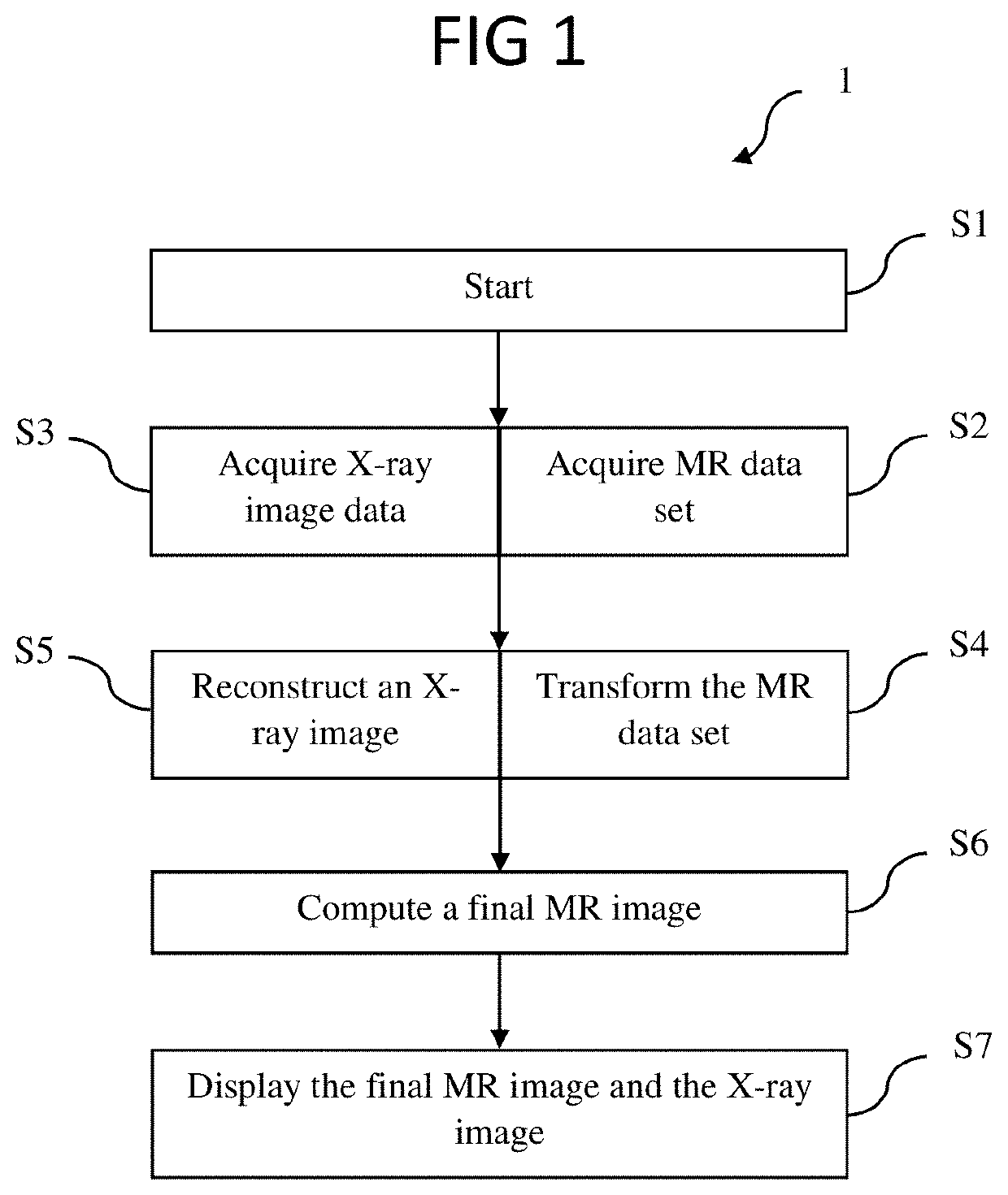
[0049]FIG. 1 schematically shows an exemplary flow chart 1 illustrating a method for combined radiography and magnetic resonance imaging. In a first process act S1, the method is started. A system used to carry out the method may be activated or set up, an object or a portion of the object to be imaged may be specified, and this specification as well as any other settings or parameters and / or control actions (e.g., for triggering the execution of the method) may be detected and / or processed. The system used to carry out the method may include an X-ray device for acquiring X-ray image data of the object in a fan- or cone-beam geometry, providing that a resulting X-ray image is a projection image generated by X-raying or radiographing the object with X-rays from a point source and detecting the X-ray radiation transmitted through the object (e.g., using an X-ray detector extending in two dimensions). The system may further include a magnetic resonance (MR) imaging device for acquiring...


PUM


Abstract

A method and a system for magnetic resonance imaging are provided. The method includes acquiring a magnetic resonance (MR) data set of an object by sampling only a portion of a k-space of the object. This sampled portion may be substantially triangle-shaped or be composed of multiple planes that extend through a point of origin of the k-space and are tangential to a common spherical cap in the k-space. An inverse Fourier transformation is then applied to the MR data set to transform the MR data set from the k-space to image space. A final MR image with a fan- or cone-beam geometry is then computed based on the transformed MR data set in image space.

Description

[0001]This application claims the benefit of EP17187933.1, filed on Aug. 25, 2017, which is hereby incorporated by reference in its entirety.BACKGROUND[0002]The present embodiments relate to magnetic resonance imaging.[0003]The activity leading to this application has received funding from the European Institute of Innovation and Technology (EIT) under grant agreement No EIT / EIT HEALTH / SGA2017 / 1. This European body receives support from the European Union's Horizon 2020 research and innovation program.[0004]Especially in medical applications, such as stroke therapy, different types of imaging devices and methods, such as X-ray imaging, magnetic resonance (MR) imaging or angiography, may be useful. It is common practice in stroke therapy to use a conventional C- arm X-ray device to acquire several projection images during an intervention to provide a physician with images of, for example, a catheter and a vessel bifurcation. These projection images may be acquired from different angu...

Claims


Application Information

Patent Timeline
Patent Type & Authority Patents(United States)
IPC IPC(8): G01R33/48G06T11/00G01R33/56A61B6/02A61B6/00G01R33/561
CPCG01R33/4824G01R33/4826G01R33/4812G01R33/482G01R33/5608G01R33/4822G01R33/4818G01R33/561G06T11/006G06T11/005A61B6/027G06T11/008A61B6/5247
Inventor LEGHISSA, MARTINOMAIER, ANDREASSTIMPEL, BERNHARDSYBEN, CHRISTOPHER
Owner SIEMENS HEALTHCARE GMBH
Why Eureka
Try Eureka