Ultrasound system and method of vessel identification

a technology of ultrasound system and detection method, applied in the field of ultrasound system and method, can solve the problems of inability to fully automatic approach robust or reliable, difficult detection of vessel walls, etc., and achieve the effect of improving ultrasound system and method for vessel detection and/or identification

Active Publication Date: 2020-07-28
KONINK PHILIPS ELECTRONICS NV
View PDF43 Cites 4 Cited by
  • Summary
  • Abstract
  • Description
  • Claims
  • Application Information

AI Technical Summary

Benefits of technology

[0006]Therefore, it would be advantageous to provide an improved ultrasound system and method for vessel detection and / or identification.
[0008]On the one hand, there is no need to manually place any seed point for vessel segmentation, and a fully automatic measurement is achieved, which reduces user dependency and enables a fast measurement. Further, a B-mode data frame is known to be useful and reliable for vessel identification, because it provides various anatomic information, such as the position of the vessel wall. On the other hand, the segmentation of the vessel in a B-mode data frame makes use of information from blood flow data frames, such as Doppler data frames, and thus more robust, reliable segmentation can be achieved. In other words, the selected blood flow region detected in data frames in a certain ultrasound mode, namely ultrasound blood flow data frame, can be served as reliable seed points for further vessel identification on basis of data frames in another ultrasound mode, namely the vessel segmentation in the B-mode data frame. Any existing or future developed, seeds-based segmentation methods can be applied to segment the vessel based on the selected blood flow region. Furthermore, the blood flow region is selected in a sequence of ultrasound blood flow data frames such as a sequence of ultrasound Doppler data frame, namely from multiple data frames acquired at different time points rather than from a single data frame. This is particularly advantageous for identifying a vessel having a blood flow which varies over time, for example, due to the cardiac cycle and / or the respiration cycle, because the blood flow of the vessel might be hardly detectable in a certain data frame but can be reliably detected in at least one data frame of the sequence. For example, the blood flow of the IVC at the expiration phase is more detectable than other phases.

Problems solved by technology

However, both are user dependent and not preferred in time critical situations, such as EMTs in pre-hospital situations.
However, the walls of the vessel are not easy to detect because other structures (tissue, bones or other vessels) may impair the detection, and so such a fully automatic approach is not robust or reliable.

Method used


Image

  • Ultrasound system and method of vessel identification
  • Ultrasound system and method of vessel identification
  • Ultrasound system and method of vessel identification

Examples


Embodiment Construction

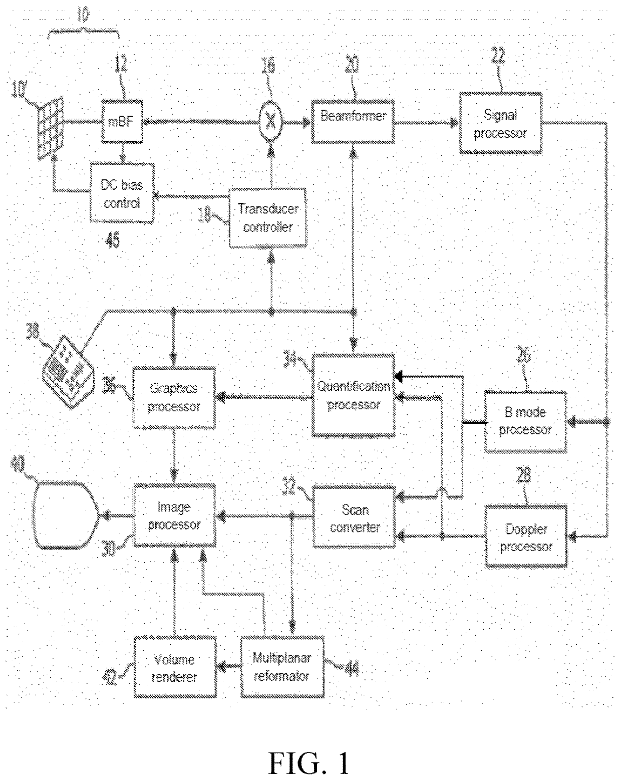
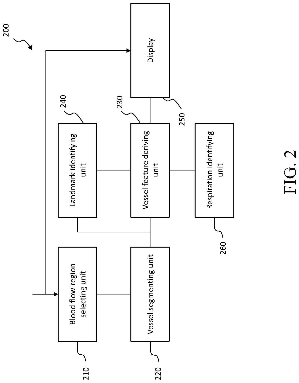
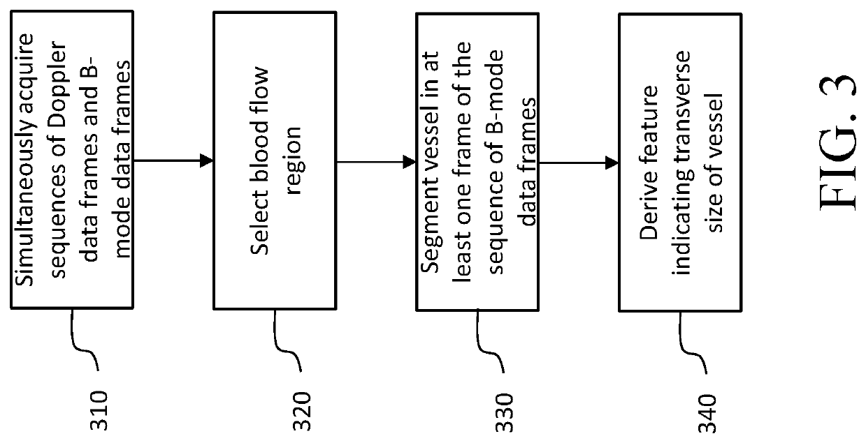
[0045]The present invention will be described with respect to particular embodiments and with reference to certain drawings but the invention is not limited thereto but only by the claims. For instance, a sequence of ultrasound Doppler data frames is taken as an example of a sequence of ultrasound blood flow data frames, but the skilled person in the art would appreciate that the sequence of ultrasound blood flow data frames can be other sequence of ultrasound data frames comprising blood flow information, such as a sequence of contrast-enhanced data frames. The drawings described are only schematic and are non-limiting. In the drawings, the size of some of the elements may be exaggerated and not drawn to scale for illustrative purposes.

[0046]Referring first to FIG. 1, an ultrasonic system with an array transducer probe is shown in block diagram form. In FIG. 1, a CMUT transducer array 10′ is provided in an ultrasound probe 10 for transmitting ultrasonic waves and receiving echo inf...


PUM

PropertyMeasurementUnit
lengthaaaaaaaaaa
ultrasoundaaaaaaaaaa
sizeaaaaaaaaaa

Abstract

An ultrasound system for identifying a vessel of a subject comprises: an ultrasound probe configured to simultaneously acquire a sequence of ultrasound blood flow data frames (such as a sequence of ultrasound Doppler data frames) and a sequence of ultrasound B-mode data frames of a region of interest including the vessel over a predetermined time period; a blood flow region selecting unit configured to select a blood flow region in the sequence of blood flow data frames; and a vessel segmenting unit configured to segment the vessel in at least one frame of the sequence of ultrasound B-mode data frames based on the selected blood flow region. Since there is no need to manually place any seed point for vessel segmentation any more, the user dependency is reduced and a fast measurement is made possible.

Description

[0001]This application is the U.S. National Phase application under 35 U.S.C. § 371 of International Application No. PCT / EP2016 / 056984, filed on Mar. 31, 2016, which claims the benefit of Chinese Application No. PCT / CN2015 / 075831, filed Apr. 3, 2015, and European Application Serial No. 15170630.6, filed Jun. 4, 2015. These applications are hereby incorporated by reference herein.FIELD OF THE INVENTION[0002]The present invention relates to an ultrasound system and method, particularly to an ultrasound system and method for vessel identification and / or the relevant measurements.BACKGROUND OF THE INVENTION[0003]Measurements of the diameter of the inferior vena cava (IVC) and its respirophasic variations, called Caval index (CI), using ultrasound have been gaining increasing importance for handling trauma.[0004]Currently, the diameter measurement of a vessel is mostly performed manually or by using semi-automatic segmentation based on seed points manually placed in the vessel region. Ho...

Claims


Application Information

Patent Timeline
Patent Type & Authority Patents(United States)
IPC IPC(8): A61B8/06A61B8/08A61B8/00A61B8/14G01B17/00G01N29/24
CPCA61B8/06A61B8/5246A61B8/488G01B17/00A61B8/5223A61B8/523A61B8/14A61B8/4494A61B8/5284A61B8/469A61B8/481A61B8/0891A61B8/085G01N2291/02466G01N2291/017G01N29/2437G01N29/2406G16H50/30
Inventor CHEN, JIANGANGRAJU, BALASUNDAR IYYAVULEYVI, EVGENIY
Owner KONINK PHILIPS ELECTRONICS NV
Why Eureka
Try Eureka