Multi-layer skin substitute products and methods of making and using the same

a skin substitute and multi-layer technology, applied in the field of live, artificial, skin substitute products and methods, can solve the problems of limited treatment options for patients, limited current commercially available skin cellular models, and insufficient recapitulation of native skin by most current engineered skins or skin substitutes

Active Publication Date: 2020-08-25
WAKE FOREST UNIV HEALTH SCI INC
View PDF32 Cites 2 Cited by
  • Summary
  • Abstract
  • Description
  • Claims
  • Application Information

AI Technical Summary

Problems solved by technology

However, due to donor-site tissue availability, complex maintenance and costs of such tissues, this treatment is often limited for patients.
Also, most current engineered skins or skin substitutes do not fully recapitulate native skin as they are devoid of multiple skin cell types and structures like trilayers and dermal appendages.
The current commercially available skin cellular models are also limited as they only use either immortalized cell lines derived from skin tumors or one or two primary cell types (e.g., keratinocytes and / or dermal fibroblasts) to be simple; thus they do not well represent and replicate the complexity of in vivo skin.

Method used


Image

  • Multi-layer skin substitute products and methods of making and using the same
  • Multi-layer skin substitute products and methods of making and using the same
  • Multi-layer skin substitute products and methods of making and using the same

Examples


examples

Fabrication of Bioprinted Skin Substitutes In Vitro

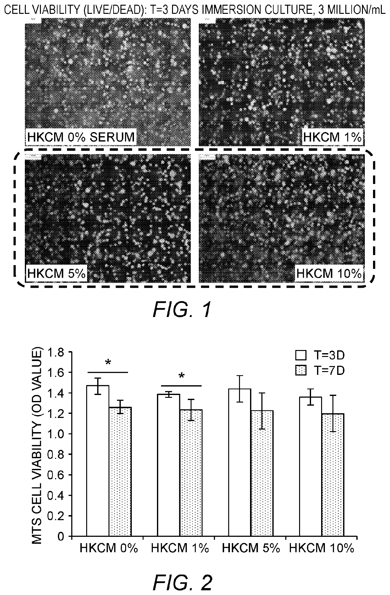
[0083]In this example, pilot studies for optimizing culture conditions for 3D reconstructed or bioprinted in vitro skin substitutes with five human primary skin cells seeded with hyaluronan-gelatin based polyethylene glycol (PEG) hydrogels are presented.

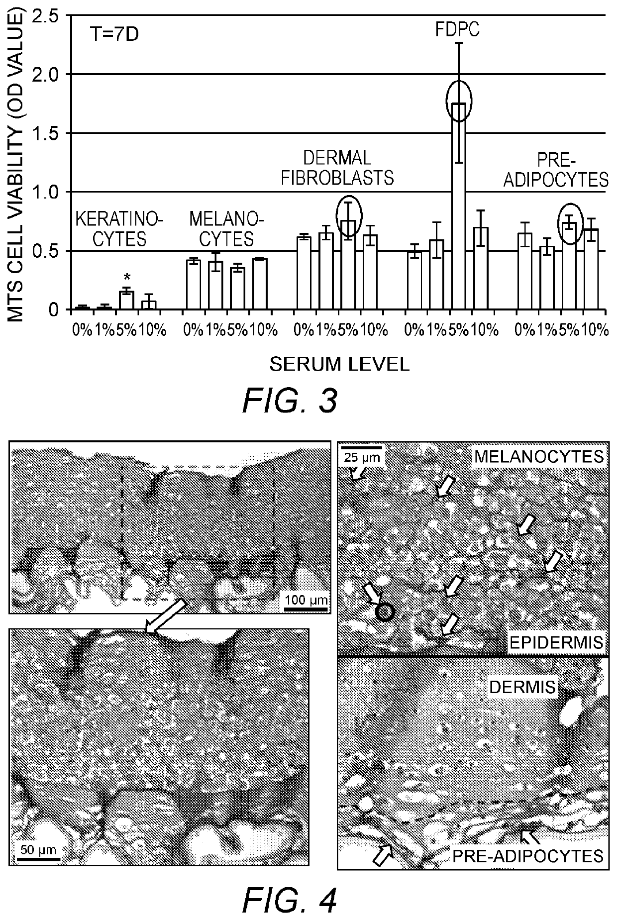
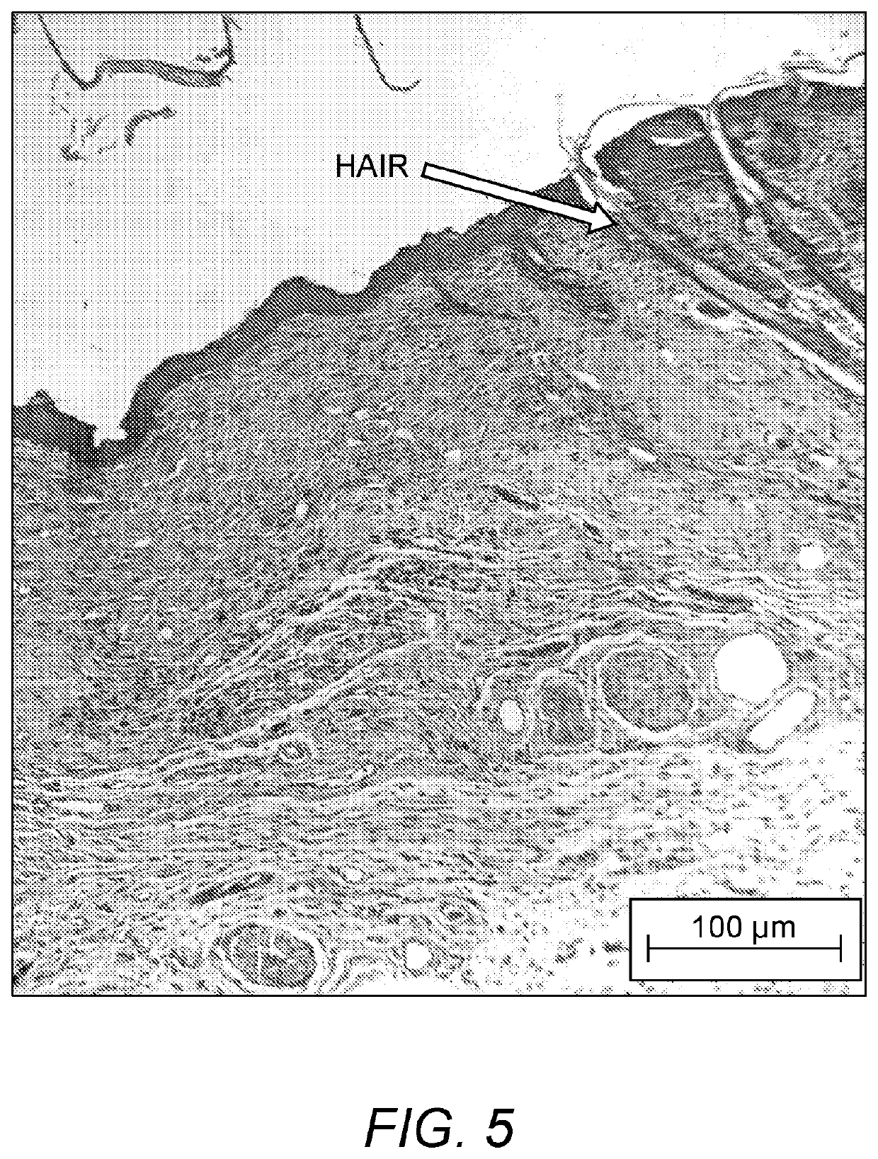
[0084]Objectives of these Examples are: [1] To test and compare the viability and proliferation of cells seeded within gels with different media conditions (0, 1, 5, & 10% serum level); [2] To test the feasibility of encapsulating 5 different skin cells within hydrogels with even distribution and to determine an appropriate cell density per construct; and [3] To show the feasibility of bioprinting trilayered 3D skin constructs in vitro with nice layering and cell presentation.

[0085]Cell Sources. Human adult keratinocytes (K), melanocytes (Mel), dermal fibroblasts (DF), follicle dermal papilla cells (FDPC), and pre-adipocytes (p-Ad) and related growth media (GM) and differentiation me...


PUM

PropertyMeasurementUnit
thicknessaaaaaaaaaa
thicknessaaaaaaaaaa
thicknessaaaaaaaaaa

Abstract

Provided are live, artificial, skin substitute products and methods of making and using the same, such as for wound treatment and compound testing, including compound testing for efficacy, toxicity, penetration, irritation and / or metabolism testing of drug candidates or compositions such as cosmetics. Described herein is an artificial mammalian skin substitute product, comprising: (a) optionally, but in some embodiments preferably, a first (“hypodermis-like”) layer comprising live mammalian adipocytes (e.g., induced pre-adipocytes) in a first hydrogel carrier; (b) a second (“dermis-like”) layer contacting or directly contacting the first layer and comprising live mammalian fibroblast cells and' live mammalian follicle dermal papilla cells in combination in a second hydrogel carrier; (c) a third (“epidermis-like”) layer contacting or directly contacting the second layer (i.e., on the opposite side thereof as the first layer, so that the second layer is sandwiched between the first and third layers when the first layer is present), the third layer comprising live mammalian keratinocytes and live mammalian melanocytes in combination in a third hydrogel carrier.

Description

RELATED APPLICATIONS[0001]This application is a 35 U.S.C. § 371 national phase entry of PCT Application PCT / US2016 / 012846, filed Jan. 11, 2016, and published in English on Jul. 21, 2016, as International Publication No. WO 2016 / 115034, and which claims the benefit of U.S. Provisional Patent Application Ser. No. 62 / 102,154, filed Jan. 12, 2015, and U.S. Provisional Patent Application Ser. No. 62 / 242,008, filed Oct. 15, 2015, the disclosure of each of which is incorporated by reference herein in its entirety.FIELD OF THE INVENTION[0002]The present invention concerns live, artificial, skin substitute products and methods of making and using the same, such as for wound treatment and compound testing.BACKGROUND OF THE INVENTION[0003]The current “gold standard” for skin replacement is the use of autologous skin grafts. However, due to donor-site tissue availability, complex maintenance and costs of such tissues, this treatment is often limited for patients. Also, most current engineered s...

Claims


Application Information

Patent Timeline
Patent Type & Authority Patents(United States)
IPC IPC(8): A61L27/60A61L27/52G01N33/50A61F2/10B33Y80/00B33Y70/00C08L5/08A61L27/20C08L89/06C12N5/071B33Y10/00B29C64/106A61L27/18A61L27/38B29K75/00B29K105/00
CPCC08L89/06A61L27/60B29C64/106B33Y80/00A61L27/20A61F2/10B33Y10/00G01N33/5044G01N33/502A61L27/3804B33Y70/00C12N5/0698A61L27/3891A61L27/52A61L27/3813A61L27/3839C08L5/08A61L27/18C12N2502/1323A61F2240/001C12N2533/80A61F2210/0076C12N2533/54B29K2075/00A61L2300/64A61L2430/34C12N2533/30B29K2105/0061C12N2502/091C12N2502/092C12N2502/1305C12N2502/094A61P17/02B29C64/112A61F2/105A61L27/24A61L27/26
Inventor ATALA, ANTHONYJEONG, GAYOUNGYOO, JAMES J.LEE, SANG JINSEOL, YOUNG-JOON
Owner WAKE FOREST UNIV HEALTH SCI INC
Why Eureka
Try Eureka