
Method for producing adenohypophysis or precursor tissue thereof

a technology of adenohypophysis and precursor tissue, which is applied in the field of producing adenohypophysis or precursor tissue thereof, can solve the problems of difficult to form hypophysis tissue from stem cells such as embryonic stem cells

Active Publication Date: 2020-09-01
RIKEN +1
View PDF6 Cites 2 Cited by
  • Summary
  • Abstract
  • Description
  • Claims
  • Application Information

AI Technical Summary

Benefits of technology

This approach successfully induces adenohypophysis or precursor tissue with the ability to regulate pituitary hormone secretion in response to hypothalamic stimulation and feedback, mirroring the body's hypophysis function in vitro.

Problems solved by technology

However, since hypophysis is formed through an extremely complicated developmental process in the embryo, it has been difficult to form hypophysis tissue from stem cells such as Embryonic Stem (ES) cells and the like.

Method used


Image

  • Method for producing adenohypophysis or precursor tissue thereof
  • Method for producing adenohypophysis or precursor tissue thereof
  • Method for producing adenohypophysis or precursor tissue thereof

Examples


example 1

[Example 1] Steric Formation of Ventral Hypothalamus and Epidermal Placode from Human Pluripotent Stem Cells

(Method)

[0146]Human ES cells (KhES-1) were subjected to maintenance culture by a conventional method on MEF and used. To monitor differentiation induction into a hypothalamus tissue, KhES-1 wherein Venus cDNA is knocked-in into the gene of Rx which is a hypothalamus neuroepithelium marker was used. To differentiate human ES cells by serum-free suspension culture of aggregate (SFEBq method), human ES cells were dispersed to single cells by enzyme by the method of Nakano et al. (Cell Stem Cell, 2012), and reaggregated in a low cell adhesive V-bottom 96 well plate (SUMITOMO BAKELITE). 5,000 cells were seeded per well, and cultured in the following differentiation medium at 5% CO2, 37° C. gfCDM medium (growth-factor-free Chemically Defined Medium; Wataya et al., PNAS, 2008)+5% KSR (Invitrogen).

[0147]With the day of seeding as day 0 of differentiation culture, 20 μM Y-27632 (ROCK i...

example 2

[Example 2] Differentiation of Hypophysial Placode and Self Organization of Rathke's Pouch

(Method)

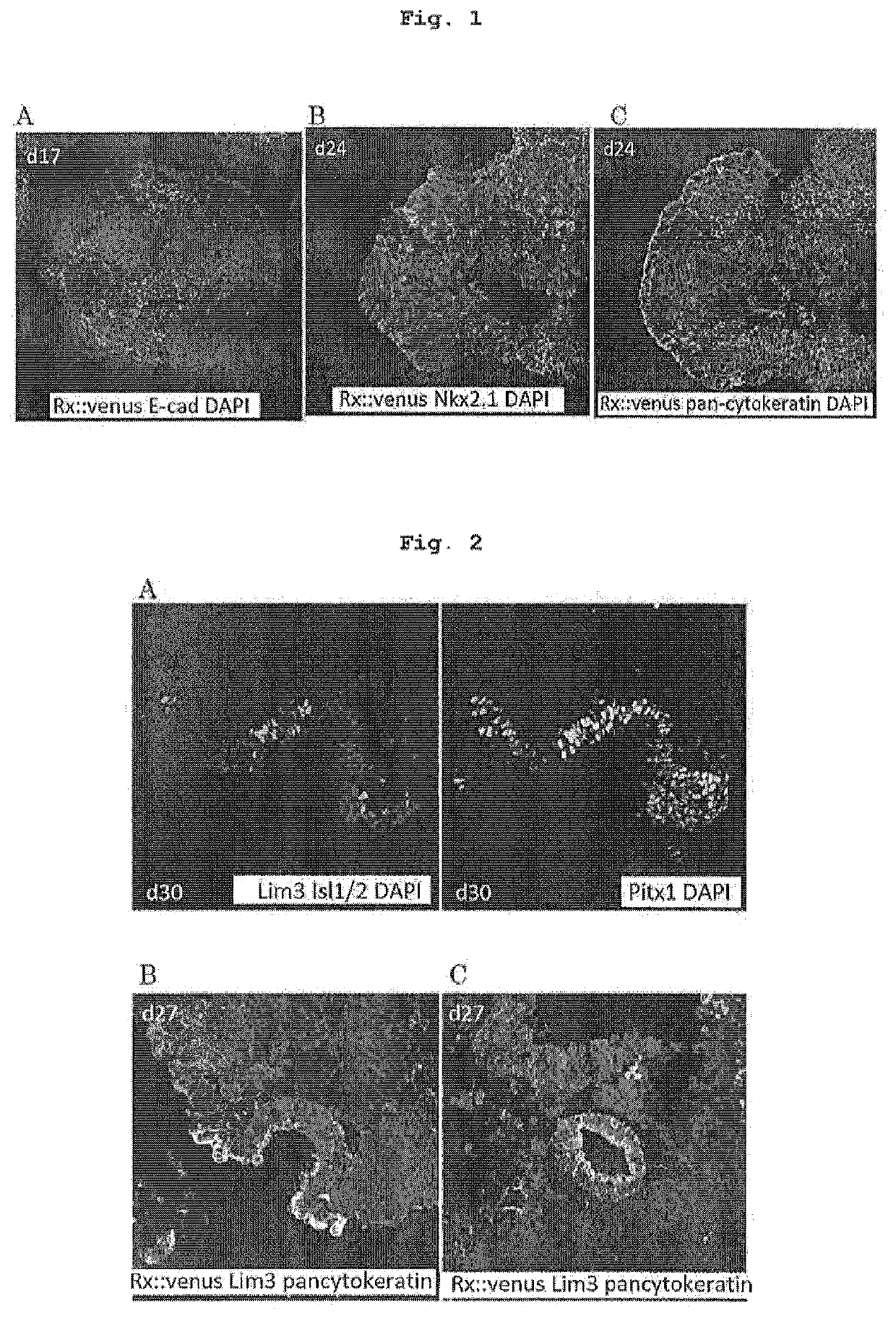
[0149]The cell aggregates derived from human ES cells and simultaneously containing ventral hypothalamus tissue and epidermal placode were formed by the method of Example 1, culture was continued until day 27 or day 30 of culture under similar conditions, and the aggregates were analyzed by a fluorescent antibody method. In some culture, FGF2 (final concentration 20 ng / ml) was added to the medium from day 15 of culture.

(Results)

[0150]On day 27 and day 30 of culture, the pan-cytokeratin-positive epidermal placode was markedly thickened and, in the analysis by a fluorescent antibody method, strongly expressed hypophysis progenitor cell markers such as Lim3, Pitx1 and Isl1 / 2 and the like (FIG. 2A). A part of the thickened placode expressing these markers was invaginated toward the inside, as in initial formation of fetal Rathke's pouch (FIG. 2B), and other part formed cyst (FIG. 2C). The a...

example 3

[Example 3] Role of BMP4 in Differentiation Induction of Hypophysial Placode

(Method)

[0151]By the method of Example 1, an influence of the presence or absence of BMP4 addition on the differentiation of cell aggregates was analyzed by a fluorescent antibody method.

(Results)

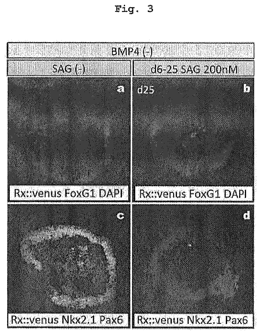
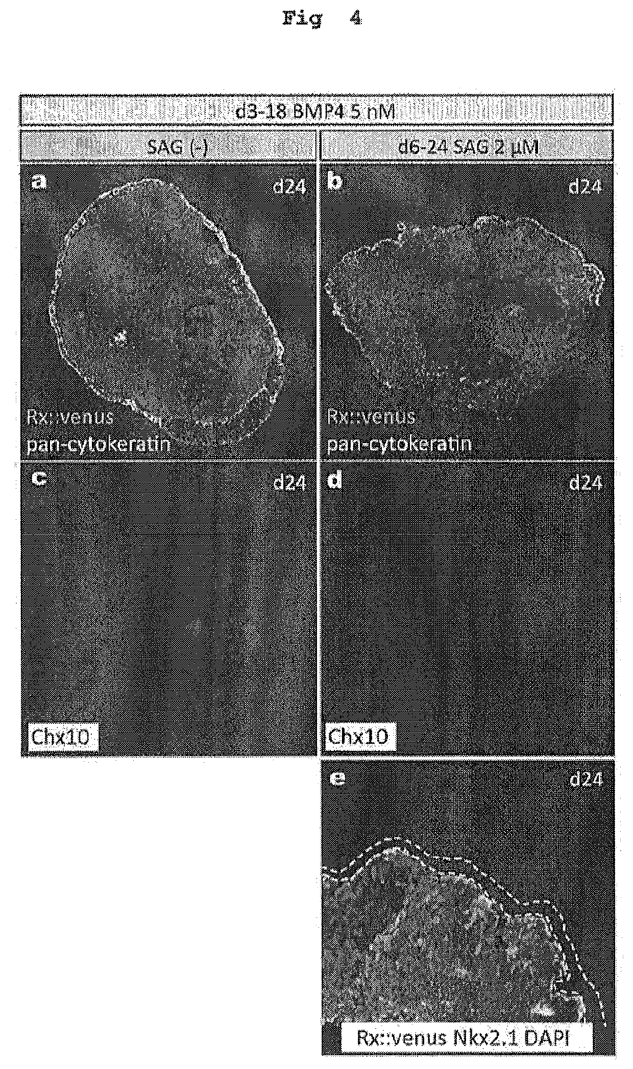
[0152]When BMP4 was not added, surface ectoderm was not formed on the surface of the aggregate on day 24 of culture, and neuroepithelium occupied the whole surface. It was found that most of the neuroepithelium did not express Rx::Venus, was FoxG1-positive and formed a cerebrum tissue (FIG. 3a, b). When SAG was not added, FoxG1-positive and Pax6-positive cerebral cortex were differentiated and when SAG was added, FoxG1-positive and Nkx2.1-positive basal ganglion was differentiated (FIG. 3a-d). Thus, suspension aggregate culture of human pluripotent stem cells has clarified that exogenous BMP4 simultaneously plays two roles of 1) active formation of surface ectoderm and 2) differentiation induction of neural tissue o...


PUM

PropertyMeasurementUnit
volumeaaaaaaaaaa
volumeaaaaaaaaaa
volumeaaaaaaaaaa

Abstract

The present invention provides a method for inducing adenohypophysis or precursor tissue thereof in vitro from human pluripotent stem cells. The method includes culturing an aggregate of human pluripotent stem cells in suspension in a medium containing a bone morphogenetic protein signal transduction pathway activating substance and a substance acting on the Shh signal pathway to obtain a human cell aggregate containing a hypothalamus neuroepithelial tissue and a surface ectoderm, and further culturing the obtained human cell aggregate containing the hypothalamus neuroepithelial tissue and the surface ectoderm in suspension in a medium containing a bone morphogenetic protein signal transduction pathway activating substance and a substance acting on the Shh signal pathway to induce formation of hypophysial placode and / or Rathke's pouch in the surface ectoderm, thus obtaining a human cell aggregate containing 1) hypothalamus neuroepithelial tissue, and 2) hypophysial placode and / or Rathke's pouch.

Description

CROSS-REFERENCE TO RELATED APPLICATIONS[0001]This patent application is the U.S. national phase of International Patent Application No. PCT / JP2015 / 071150, filed on Jul. 24, 2015, which claims the benefit of Japanese Patent Application No. 2014-152384, filed on Jul. 25, 2014, the disclosures of which are incorporated herein by reference in their entireties for all purposes.TECHNICAL FIELD[0002]The present invention relates to a technique for inducing differentiation of pluripotent stem cells into an adenohypophysis or a progenitor tissue thereof in vitro.BACKGROUND ART[0003]Hypophysis is a small endocrine organ adjacent to the lower part of the diencephalon and plays a major role as a control center of various hormones. For example, it produces various pituitary hormones including adrenocorticotropic hormone (ACTH) which promotes production of corticosteroid essential for life support, growth hormone which promotes growth of children, and the like. Therefore, functional disorder of h...

Claims


Application Information

Patent Timeline
Patent Type & Authority Patents(United States)
IPC IPC(8): C12N5/00C12N5/079C12N5/0735C12N5/02
CPCC12N5/0606C12N5/0037C12N5/0031C12N5/0618C12N2501/41C12N2501/42C12N2501/115C12N5/0616C12N2501/155C12N2506/02C12N2506/45
Inventor SASAI, YOSHIKIOZONE, CHIKAFUMISUGA, HIDETAKA
Owner RIKEN
Learn More