Automatic device-footprint-free roadmapping for endovascular interventions

a technology of endovascular interventions and automatic devices, applied in the field of endovascular intervention road mapping, can solve the problems of affecting the visualization effect of endovascular interventions, so as to achieve enhanced roadmapping visualization

Active Publication Date: 2020-11-03
KONINKLJIJKE PHILIPS NV
View PDF16 Cites 0 Cited by
  • Summary
  • Abstract
  • Description
  • Claims
  • Application Information

AI Technical Summary

Benefits of technology

[0009]Thus, there may be a need to provide a system and a method which allows for an enhanced roadmapping visualization during endovascular interventions.
[0010]According to a first aspect of the present invention an imaging system for automatic roadmapping for endovascular interventions is provided. The system comprises an x-ray imaging device for acquiring x-ray images and a calculation unit. The x-ray imaging device is adapted for acquiring a first x-ray image with an interventional device present in the vessels while no contrast agent is injected into the vessels. Furthermore, the x-ray imaging device is adapted for acquiring a second x-ray image with the interventional device present in the vessels while contrast agent is injected into the vessels. The calculation unit is adapted for creating a roadmap image by subtracting the first x-ray image from the second x-ray image. Moreover, the calculation unit is adapted for automatically minimizing the visibility of the interventional device in the roadmap image.
[0011]In other words, the idea of the invention may be seen in providing a roadmap and possibly also a visualization of a roadmap that removes explicitly the devices footprint during the mask image generation. This allows for obtaining a useable roadmap when there is some motion of objects in the acquired images. In a first approach, for enhancing the quality of the roadmap image the footprint of the interventional device may automatically be detected in injected and non-injected frames of the angiogram or aortogram. Subsequently, the interventional device may be in-painted in each frame before generating the roadmap image. In a second approach, after subtracting the injected and non-injected frames from each other, the interventional device may be removed by filtering and saturating the roadmap image. In both approaches, the resulting roadmap image does not contain anymore the footprint of the interventional device, such that the treatment area is optimally visible.

Problems solved by technology

The result may then be overlaid on live fluoroscopic frames bringing the vessels footprint on live images.
However, when there is some motion, for instance due to breathing, the mask image may contain a lot of artefacts.
In particular, the artefacts may be caused by interventional devices present in the vessels during image acquisition.
The resulting overlaid image may be not very helpful.
When the mask image is overlaid on fluoroscopic frames the device may appear three times. Moreover, those artefacts may often be located at the treatment area and may therefore occlude the visualization of the devices in the live overlaid frame.
However, this restricts the acquisition protocols and / or dosage for angiograms and fluoroscopy, because the same acquisition protocols and / or dosage have to be used for both image sequences.

Method used


Image

  • Automatic device-footprint-free roadmapping for endovascular interventions
  • Automatic device-footprint-free roadmapping for endovascular interventions
  • Automatic device-footprint-free roadmapping for endovascular interventions

Examples


Embodiment Construction

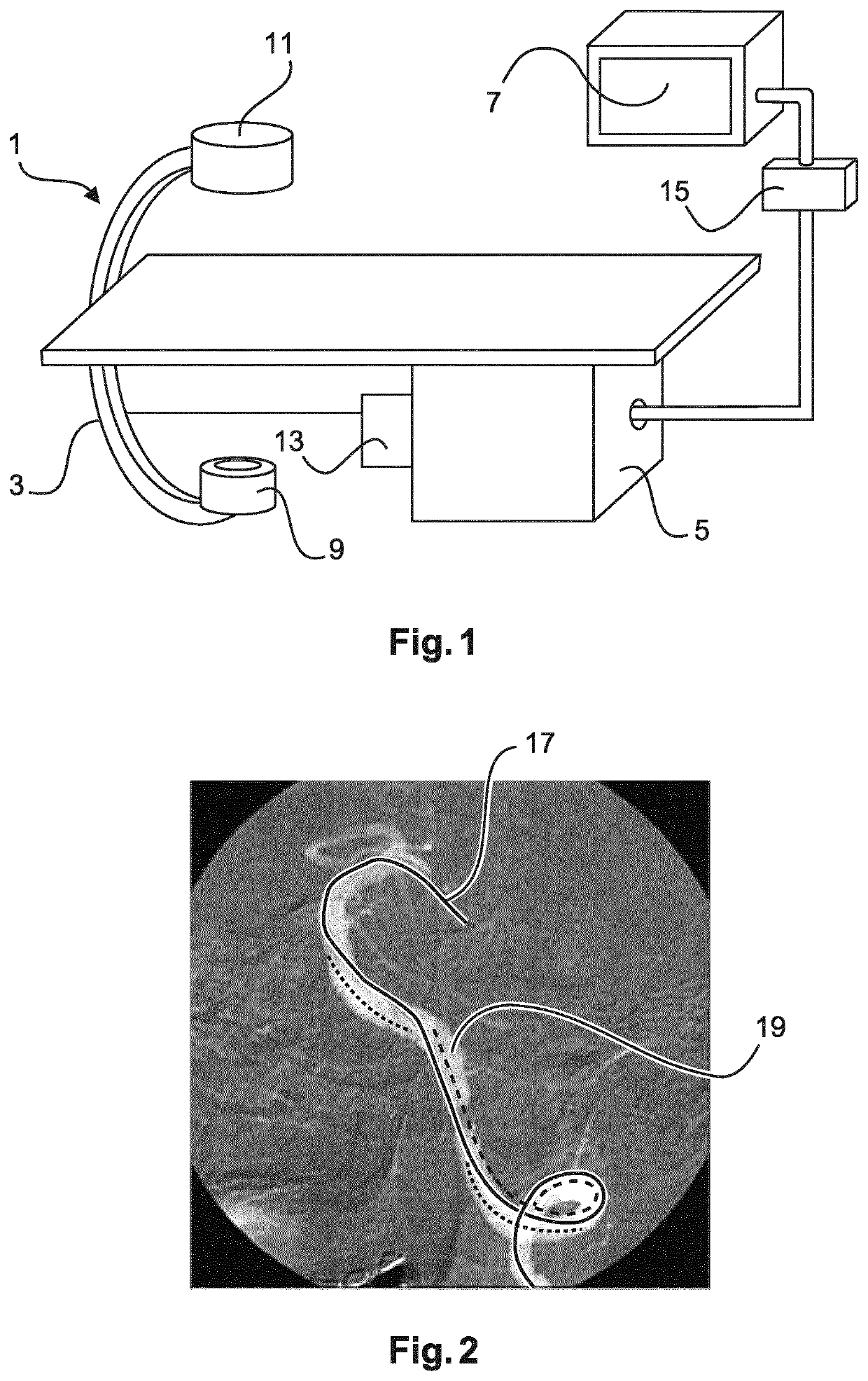
[0041]FIG. 1 schematically shows an imaging system 1 for automatic roadmapping for endovascular interventions. For example, the system 1 may be employed in a catheterization laboratory. The system 1 comprises an x-ray imaging device 3 for acquiring x-ray images. The imaging device 3 may comprise an x-ray radiation source 9 and an x-ray detection module 11. The x-ray detection module 11 may be positioned opposite the x-ray radiation source 9. During the examination and / or intervention procedure a subject such as a patient is located between the x-ray detection module 11 and the x-ray radiation source 9, e.g. on a table.

[0042]Furthermore, the imaging system 1 comprises a calculation unit 5 which may for example be a processor unit with a memory unit 13. Therein, the calculation unit 5 is adapted to execute an algorithm which may for example be stored on the memory unit 13. The calculation unit 5 is electrically and functionally connected to the x-ray imaging device 3.

[0043]Moreover, t...


PUM


Abstract

A system (1) and a corresponding method enable an enhanced roadmapping visualization without unnecessary device-footprints. The system (1) includes an x-ray imaging device (3) for acquiring x-ray images and a calculation unit (5). The x-ray imaging device (3) is adapted for acquiring a first x-ray image (21) with an interventional device (17) present in the vessels (19) while no contrast agent is present in the vessels (19) and a second x-ray image (23) with the interventional device (17) present in the vessels (19) while contrast agent is present in the vessels (19). The calculation unit is adapted for creating a roadmap image (27) by subtracting the first x-ray image (21) from the second x-ray image (23) and automatically minimizing the visibility of the interventional device (17) in the roadmap image (27). A display unit (7) is adapted to display the roadmap image (27) or an overlay of a current fluoroscopy image (31) with the roadmap image (27).

Description

CROSS-REFERENCE TO PRIOR APPLICATIONS[0001]This application is the U.S. National Phase application under 35 U.S.C. § 371 of International Application No. PCT / EP2014 / 069144, filed on Sep. 9, 2014, which claims the benefit of European Patent Application No. 13306286.9, filed on Sep. 20, 2013. These applications are hereby incorporated by reference herein.FIELD OF THE INVENTION[0002]The present invention relates to roadmapping for endovascular interventions. In particular, the present invention relates to a system and a corresponding method for automatic roadmapping for endovascular interventions. Furthermore, the present invention relates to a computer program element and a computer readable medium.BACKGROUND OF THE INVENTION[0003]During endovascular procedures under x-ray guidance such as Endovascular Aneurysm Repair (EVAR) catheters and treatment devices are inserted in the vasculature though a small incision e.g. in the abdominal region. The user then moves the tools to the treatme...

Claims


Application Information

Patent Timeline
Patent Type & Authority Patents(United States)
IPC IPC(8): A61B6/00A61B6/12A61B6/04
CPCA61B6/461A61B6/12A61B6/487A61B6/5235A61B6/481A61B6/5252A61B6/504A61B6/4441A61B6/463A61B6/0407
Inventor NEMPONT, OLIVIER PIERRECATHIER, PASCAL YVES FRANCOISFLORENT, RAOULPIZAINE, GUILLAUME JULIEN JOSEPH
Owner KONINKLJIJKE PHILIPS NV
Why Eureka
Try Eureka