
Systems and methods for diagnosis of middle ear conditions and detection of analytes in the tympanic membrane

a technology of tympanic membrane and middle ear, applied in the field of otolaryngology, can solve the problems of difficult to make a rapid and accurate many patients being over-treated for their condition, and difficulty in reliable diagnosis of middle ear conditions

Active Publication Date: 2020-12-29
CONNECTICUT CHILDRENS MEDICAL CENT +1
View PDF39 Cites 1 Cited by
  • Summary
  • Abstract
  • Description
  • Claims
  • Application Information

AI Technical Summary

Benefits of technology

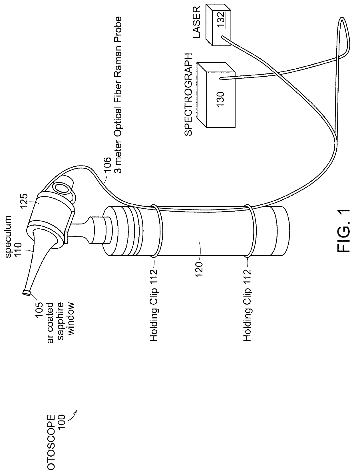
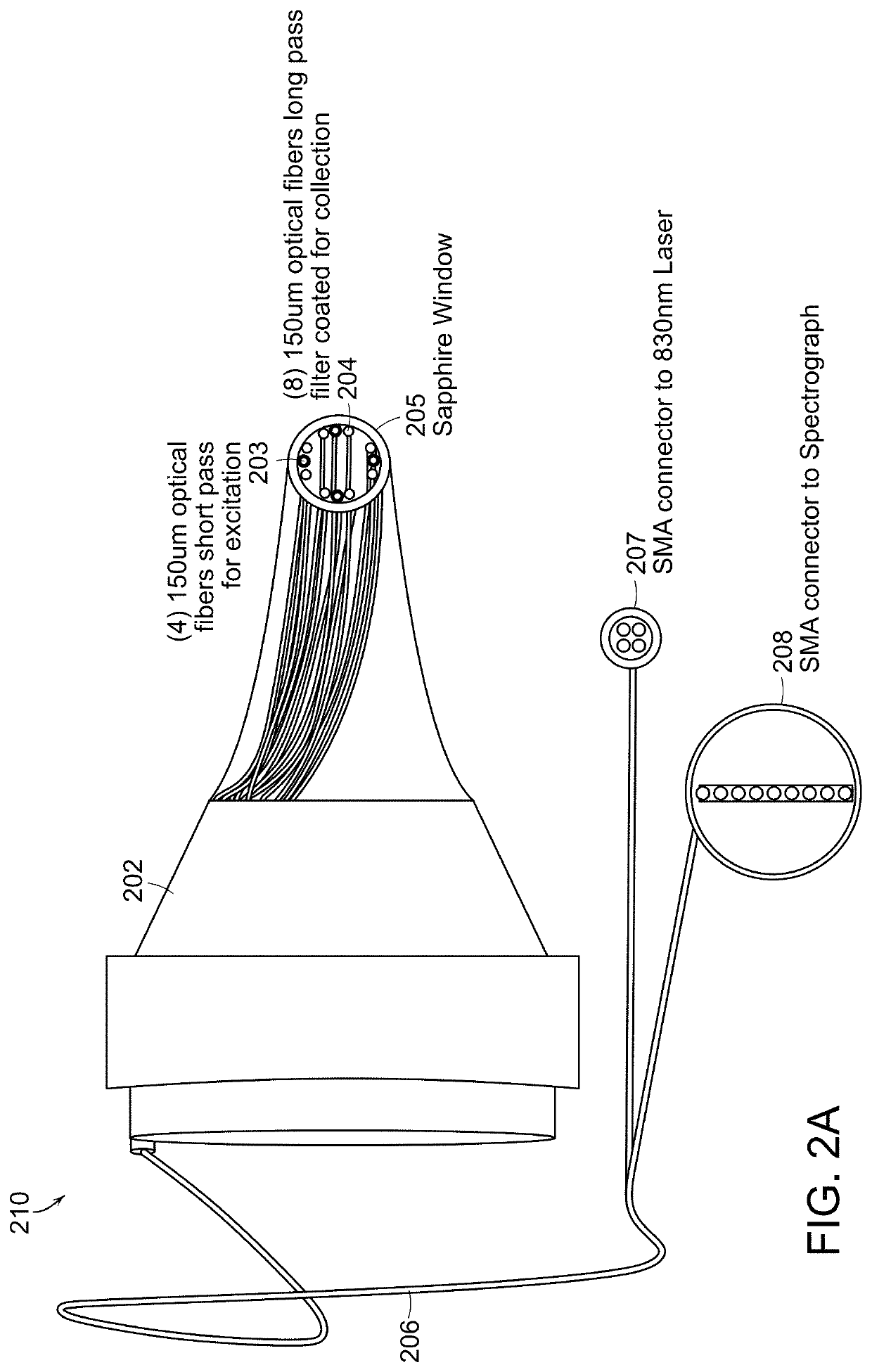
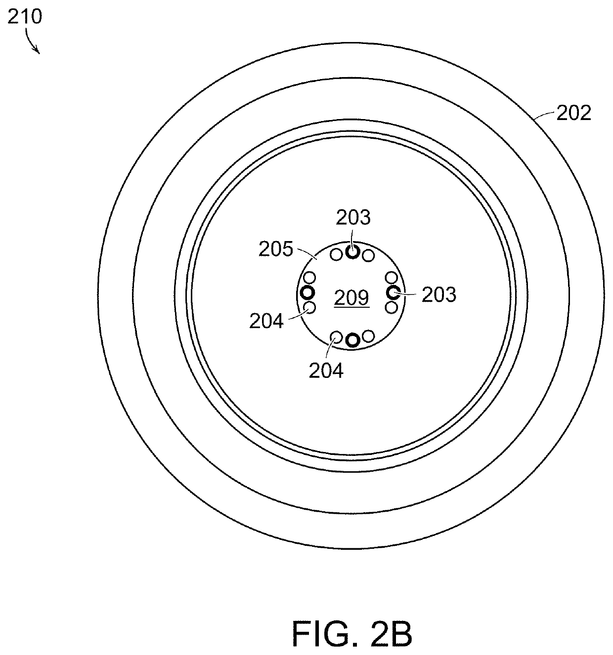
The present invention is about devices and methods for measuring and imaging middle ear pathologies using spectral analysis. This technology can help diagnose and treat common ear conditions like cholesteatoma, which is a proliferative lesion that can damage the hearing. The device is a low-cost, detailed and objective tool that can provide valuable information to guide the surgical procedure and reduce the need for revision surgeries. The invention uses an existing otoscope platform and incorporates optical components as a self-contained feature that can also be used in an otoendoscope. The device design allows for concomitant fluorescence measurements and conventional otoscopic evaluation to reduce registration errors. Overall, this technology provides a novel mechanism to acquire real-time images of the tissue epithelium, optimizing the value of the initial procedure and reducing the necessity for revision surgeries.

Problems solved by technology

A common problem in otolaryngology is the lack of certainty regarding diagnosis for middle ear conditions resulting in many patients being over-treated for their condition.
Despite the high incidence of clinical visits that pertain to middle ear pathologies, the ability to make a rapid and accurate diagnosis of these conditions has not improved considerably.
Reliable diagnosis of middle ear conditions remains challenging, and the accuracy of currently available methods is limited.
Nevertheless, they represent a surgical condition, and resolution of the disease process can only occur after complete externalization or removal of the lesion.
However, this may require multiple surgical procedures that can create significant burdens for those affected by this disease.
However, classical otoscopic evaluation suffers from observer variability and provides limited insight into a disease's defining biochemistry.
While relatively accurate prediction has been achieved in the past, this success has been limited to a personalized setting because skin tissue varies highly from one person to another and a single set of calibration data cannot be used across multiple patients.
1. Interference from unwanted optical signals arising from dermis and epidermis tissues and from tissue turbidity.
2. Measured Raman signals from analytes are detected mostly from the interstitial fluid (ISF) and not from the actual blood as the light sampling volume for wavelengths typically used (including near infrared wavelengths) is quite limited. There is a known physiological lag between ISF glucose and blood glucose and, unfortunately, calibration is only possible using blood glucose. This inconsistency results in inaccuracy in the measurement.
3. Skin tissue is highly variable from one individual to another, which necessitates that measurements are person-specific which further contributes to the complexity of calibration.

Method used


Image

  • Systems and methods for diagnosis of middle ear conditions and detection of analytes in the tympanic membrane
  • Systems and methods for diagnosis of middle ear conditions and detection of analytes in the tympanic membrane
  • Systems and methods for diagnosis of middle ear conditions and detection of analytes in the tympanic membrane

Examples


Embodiment Construction

[0038]Standard otoscopic evaluation currently requires subjective visual interpretation of white light reflecting off the tympanic membrane and the middle ear promontory. In other words, the entire process of examining the normal structure, diagnosing diseases, and providing critical input for therapy relies extensively on human recognition of morphologic patterns in vivo. This approach, which is the basis for current decision-making, has been shown to be less than ideal for a number of conditions in particular when it relates to ear examination. More specifically, congenital cholesteatomas tend to be asymptomatic until they reach a significant size, and successful outcomes rely on an astute clinician to make a diagnosis before the lesion causes significant damage to middle ear structures. Detection of congenital cholesteatomas with simple otoscopic examination can be missed altogether or confused with normal conditions such as myringosclerosis. Investigators have opined that even o...


PUM

PropertyMeasurementUnit
wavelengthaaaaaaaaaa
excitation wavelengthaaaaaaaaaa
diameteraaaaaaaaaa

Abstract

Systems and methods are presented for the diagnosis of middle ear pathological conditions based on spectral signatures. Preferred embodiments provide for detection of one or more analytes from the tympanic membrane. Devices use spectral measurements including spectral imaging to non-invasively identify middle ear pathological conditions including cholesteatoma and acute otitis media by providing real-time information of differentially expressed molecules. Devices and methods can also be used to non-invasively detect and quantify blood analytes from the tympanic membrane.

Description

RELATED APPLICATION[0001]The present application claims priority to United States Provisional Patent Application 62 / 218,989, filed Sep. 15, 2015, the entire application being incorporated herein by reference.STATEMENT REGARDING FEDERALLY SPONSORED RESEARCH OR DEVELOPMENT[0002]This invention was made with Government support under Grant No. P41 EB015871 awarded by the National Institutes of Health. The Government has certain rights in the invention.BACKGROUND OF THE INVENTION[0003]A common problem in otolaryngology is the lack of certainty regarding diagnosis for middle ear conditions resulting in many patients being over-treated for their condition. While pneumatic otoscopy and adjunctive tests offer additional information, white light otoscopy has been the main tool for diagnosis of external auditory canal and middle ear pathologies for over a century.[0004]Despite the high incidence of clinical visits that pertain to middle ear pathologies, the ability to make a rapid and accurate ...

Claims


Application Information

Patent Timeline
Patent Type & Authority Patents(United States)
IPC IPC(8): A61B5/00A61B1/227A61B1/00A61B5/145A61B1/04A61B1/32A61B5/1459G01N21/65G01J3/44A61B1/07A61B5/12A61B1/06G01J3/02G01N21/64
CPCA61B5/6817G01N21/65A61B5/7282A61B1/0638G01J3/4406G01J3/0218A61B1/32A61B1/0684A61B5/14546A61B1/07A61B5/12A61B1/00045G01J3/44A61B1/227A61B5/14532A61B5/1459A61B1/043A61B1/00009A61B5/0075A61B5/0084G01N21/6486A61B5/0071A61B1/000094
Inventor PANDEY, RISHIKESHSPEGAZZINI, NICOLASGALINDO, LUIS H.DASARI, RAMACHANDRAVALDEZ, TULIO ALBERTO
Owner CONNECTICUT CHILDRENS MEDICAL CENT
Learn More