Detachable miniature microscope mounted keratometer for cataract surgery

a miniature microscope and keratometer technology, applied in the field of diagnostic instruments, can solve the problems of poor cataract surgery outcome, insufficient biometry accuracy, lack of intraoperative astigmatism measurement tools, etc., and achieve the effect of improving iol imaging and cataract visualization

Active Publication Date: 2021-01-12
ARTSYUKHOVICH ALEX
View PDF15 Cites 0 Cited by
  • Summary
  • Abstract
  • Description
  • Claims
  • Application Information

AI Technical Summary

Benefits of technology

[0011]In one embodiment, the present invention provides a keratometer for intra-surgery measurements mounted under a surgical microscope. The keratometer includes a Placido ring illuminating a patient's eye; a video camera; a beamsplitter directing a Purkinje image of the Placido ring to the video camera; a fixation light directing a beam for patient eye fixation, fixation confirmation or creating a red reflex effect to enhance IOL imaging and cataract visualization; a processor configured to determine the refractive characteristics and keratometer parameters of the patient's eye and to execute a digital image enhancement method to outline IOL features to help IOL alignment; and a digital display displaying the keratometer parameters and surgical guidance information for a surgeon.
[0017]In another embodiment, the near-collimated visible or NIR light source, when operated in a constant “ON” mode, creates a fundus reflection effect for video contrast.
[0018]In another embodiment, the digital image enhancement method is applied to live video to outline IOL features for IOL alignment ease.
[0028]In another embodiment, the keratometry method further includes providing a near-collimated visible or NIR light source to fix the patient's eye and creating a red reflex effect enhancing the contrast of an IOL or assisting cataract visualization.

Problems solved by technology

Currently insufficient accuracy of biometry results in poor cataract surgery outcomes 50% of the time, when patients have >0.5 Diopter Refractive error surgical outcome and need to wear glasses after surgery.
The lack of tools for intraoperative astigmatism measurement slows down adoption of toric IOLs that correct astigmatism.
One difficulty is to apply pre-surgery biometry that is normally done two weeks before surgery to intra-surgery guidance.
The extra step of image registration adds here an error to original pre-operative astigmatism angle measurement.
Also it is unable to accurately account for Surgically Induced Astigmatism (SIA) created post-measurement due to corneal incisions during surgery.
These are very complex and expensive optical devices that are large in size and take up to 30% of surgical space under microscope during cataract micro-surgery.
Several microscope-mounted keratometers were proposed, but none of them has been realized in commercial product.
This is due to the difficulty of achieving high accuracy in a small microscope-mounted device—the device's size is restricted by surgical space available.

Method used


Image

  • Detachable miniature microscope mounted keratometer for cataract surgery
  • Detachable miniature microscope mounted keratometer for cataract surgery
  • Detachable miniature microscope mounted keratometer for cataract surgery

Examples


Embodiment Construction

[0049]Reference will now be made in detail to embodiments of the present invention, example of which is illustrated in the accompanying drawings.

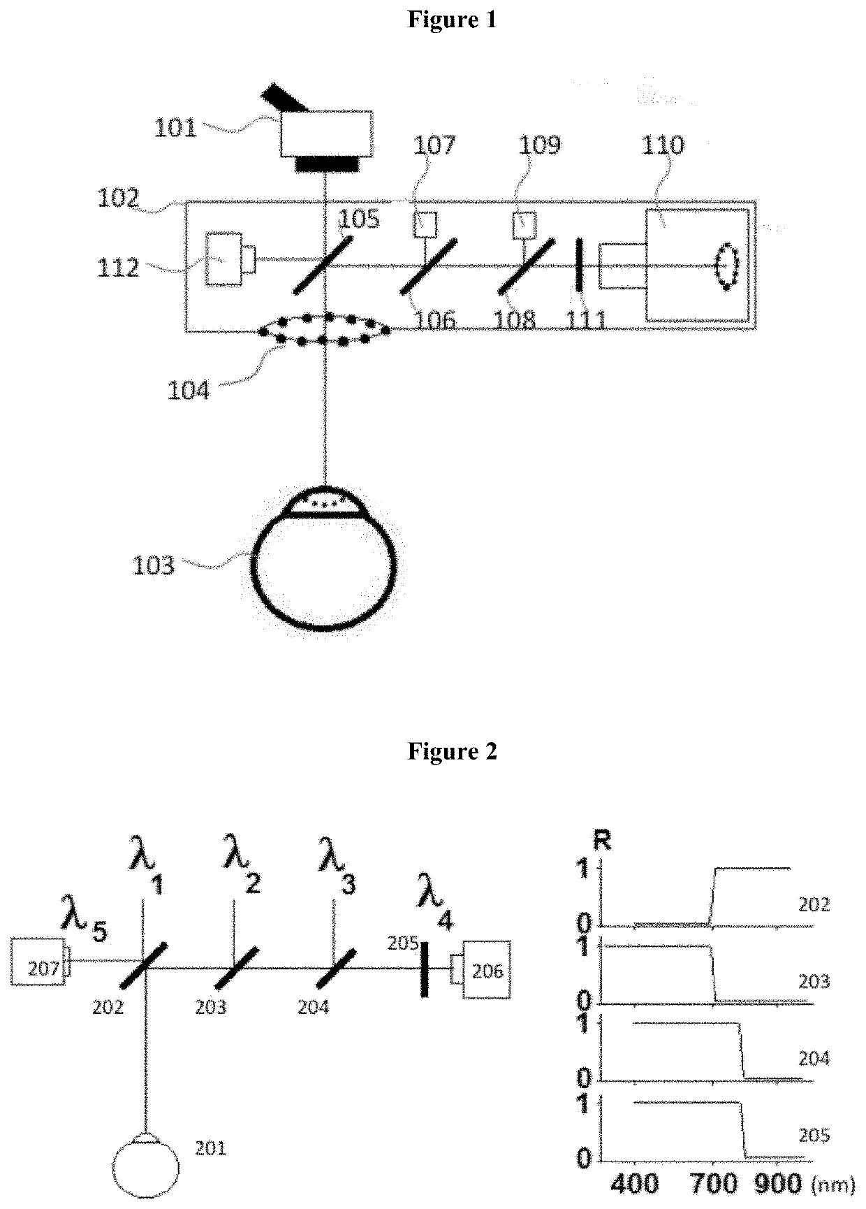
[0050]FIG. 1 depicts schematically one proposed implementation of suggested detachable miniature keratometer device. A surgical microscope 101 has the keratometer device 102 attached at the bottom of it—above a patient's eye 103. The keratometer device 102 has an illuminated Placido ring 104 (or multiple Placido rings) attached at the bottom. FIG. 1 shows it as a ring formed by 12 discrete light sources—LEDs. But one skilled in art recognizes that other means of Placido ring can be utilized—a continuous illuminated ring based on fiber optics or scattering optical material. The number of discrete light sources can be changed. One or multiple Placido rings can be projected by using a projection lens and OLED display or reticle, illuminated with light source. A Purkinje-1 image of Placido ring 104 formed on anterior cornea of the patient's eye...


PUM


Abstract

A keratometer for intra-surgery measurements mounted under a surgical microscope includes a Placido ring illuminating a patient's eye; a video camera; a beamsplitter directing a Purkinje image of the Placido ring to the video camera; a fixation light directing a beam for patient eye fixation, fixation confirmation or creating a red reflex effect to enhance IOL imaging and cataract visualization; a processor configured to determine the refractive characteristics and keratometer parameters of the patient's eye and to execute a digital image enhancement method to outline IOL features to help IOL alignment; and a digital display displaying the keratometer parameters and surgical guidance information for a surgeon.

Description

[0001]This application is the national stage application of PCT / US2017 / 028215, filed on Apr. 19, 2017, which claims priority to U.S. Provisional Application No. 62 / 391,343, filed Apr. 28, 2016, which is incorporated by reference for all purposes as if fully set forth herein.FIELD OF THE INVENTION[0002]The present invention relates to a diagnostic instrument and its application, more particularly, to a detachable keratometer for intra-surgery measurements mounted under a surgical microscope and a method of using the same.BACKGROUND OF THE INVENTION[0003]Cataract surgery is #1 in volume surgery in the World. Currently there are 3 million cataract surgeries performed each year in US. Western Europe also performs 3 million cataract surgeries. The WHO estimates that by 2020, 32 million cataract surgeries will be performed, up from 12 million in 2000. This is not just due to increased life span and baby boomer demographic population increase. More people using digital devices (tablets, sm...

Claims


Application Information

Patent Timeline
Patent Type & Authority Patents(United States)
IPC IPC(8): A61B3/10A61B3/14A61B3/02A61B3/00A61F9/007A61B3/107A61B3/13A61F2/16A61B3/036A61B90/00
CPCA61F9/00736A61B3/0091A61B3/102A61B3/107A61B3/13A61B3/145A61F2/1662A61B3/036A61B2090/3735G02B21/0012G02B21/084G02B27/1006
Inventor ARTSYUKHOVICH, ALEX
Owner ARTSYUKHOVICH ALEX
Why Eureka
Try Eureka