Motion-adaptive interactive imaging method

an interactive imaging and motion-adaptive technology, applied in the field of motion-adaptive interactive imaging methods, can solve the problems of difficult to implement margin assessments based on frozen sectioning on breast cancer specimens, difficult manipulation and orientation of complex three-dimensional (3d) primary specimens for full inspection and processing, and significant ambient interference, etc., to achieve enhanced image quality, better sensitivity, and longer integration time

Active Publication Date: 2021-05-04
LI-COR BIOTECH LLC
View PDF156 Cites 1 Cited by
  • Summary
  • Abstract
  • Description
  • Claims
  • Application Information

AI Technical Summary

Benefits of technology

[0008]In general, provided herein are imaging technology methods, computer programs, and systems for viewing a subject from multiple angles or orientations while adapting in real-time the imaging quality and imaging function in response to motion. The provided methods and systems maintain the capability of providing a real-time preview during image-guided navigation, while also including high-sensitivity image properties when an adaptive function is turned on. Motion-sensitive feedback from, for example, the movement of an imager, is used to assess user-controlled motion commands. When motion is substantially reduced or stopped, the imager is switched to an adaptive or enhanced configuration that can include additional imaging modalities, enhanced image quality, longer integration time for a better sensitivity, overlapping of channels, noise filtering, color changes, computational results, or other alterations. In this way, the adaptive imaging configuration can allow for better detection of faint signals at the location of interest that would otherwise be difficult to appreciate with the original imaging properties in use during the real-time preview and prior to the motion-adaptive switch.
[0009]For example, the capability to interactively review a sample in a 3D real-time image, with an optimal fluorescence imaging sensitivity adaptively triggered in response to a change in motion, has great potential for providing important information for the vetting of surgical samples. Furthermore, adaptive functional imaging with modalities of X-ray and high-sensitivity fluorescence imaging can significantly increase the efficiency, reduce the sampling error, and increase the diagnosis accuracy of intraoperative margin assessment in cancer surgery. Similarly, the utility of a biopsy imager can be improved by providing an interactive preview of a specimen with useful additional information enabled by adaptive imaging modalities.

Problems solved by technology

However, complex three-dimensional (3D) primary specimens are often difficult to manipulate and orient for full inspection and processing.
As a specific example, margin assessments based on frozen sectioning is difficult to implement on breast cancer specimens due to fatty tissues.
When used in situ with an image-guided systems above the wound bed, there exist significant ambient interferences such as those associated with broadband ambient light, wound bed fluids, and electro-magnetism.
While each of these can positively influence the sensitivity of fluorescence imaging, they can also negatively influence the video-rate performance of image-guided systems.
However, the typical performance characteristics of these overlays of reflective light and fluorescence images sacrifices real-time motion sensitivity as a compromise to boost fluorescence sensitivity.
In general, improvements to high frame rate imaging negatively impact the sensitivity of the imaging system by reducing signal collection time in each frame as well as by reducing data processing time and increasing read noises of the imaging sensor.

Method used


Image

  • Motion-adaptive interactive imaging method
  • Motion-adaptive interactive imaging method
  • Motion-adaptive interactive imaging method

Examples


Embodiment Construction

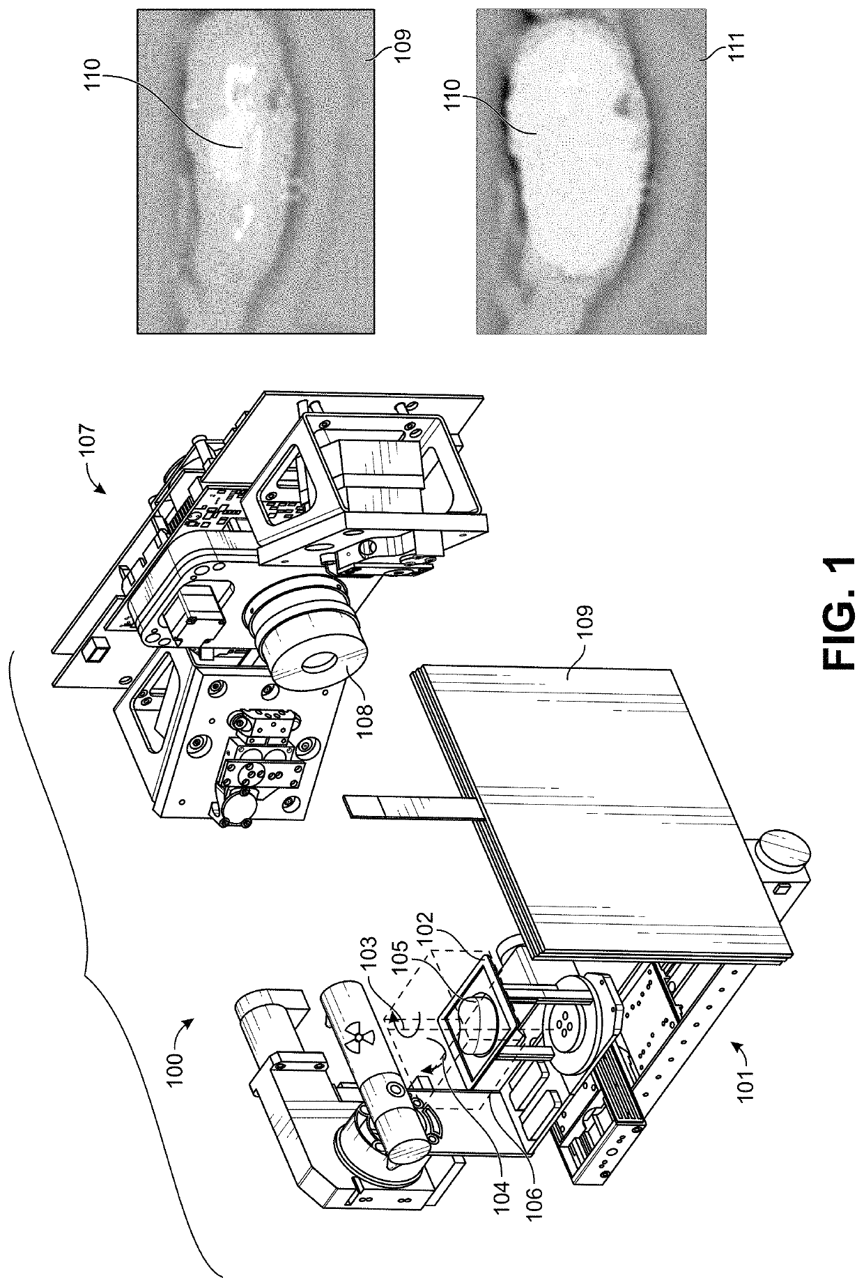
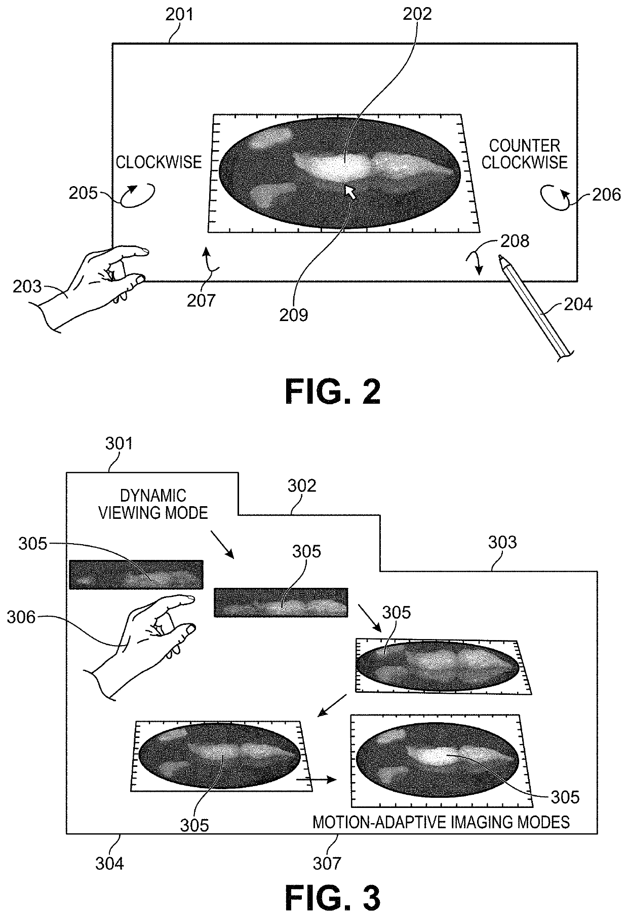
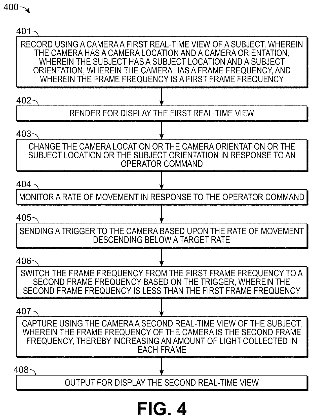
[0029]Embodiments of the present invention relate in part to motion-adaptive switching of imaging functions, such as those, for example, used for surgical or biopsy imaging. The provided methods and systems can monitor for changes in motion, and responsively enhance or modify real-time images being captured or rendered for display. This can be accomplished by analyzing user control behaviors to determine when and how to optimize imaging results, while simultaneously responding to inputs from the user who is interactively directing a viewing motion around areas of interest. In brief, the user can use motion control to direct real-time 3-D imaging of a sample. The motion-adaptive imaging can then maintain a real-time, high-speed preview as the image is in motion, while automatically switching to an enhanced functional image when the image is relatively stationary. Because the imaging needs during movement and when stationary are often different, or even contradictory, the ability to a...


PUM


Abstract

Methods and systems are provided for the detecting and presenting of faint signals from a real-time interactive imager. A real-time view of a subject is controlled by a user by entering commands that move the subject or a camera used to record the real-time view. The rate of this movement is monitored, and when the rate descends below a preset value, a trigger is sent to the camera. In response to the trigger, the camera switches to a lower frame frequency for capturing the real-time view of the subject, thereby increasing the amount of light collected in each frame, and increasing the sensitivity of the camera to faint signals. The methods and systems are particularly relevant for surgical or biopsy imaging, in which both a higher frame rate preview image of a subject for 3D visualization, and a higher sensitivity fluorescence image of the subject for tumor margin assessment, are desired.

Description

CROSS-REFERENCES TO RELATED APPLICATIONS[0001]The present application claims the benefit of U.S. Provisional Patent Application No. 62 / 425,967, filed Nov. 23, 2016, and U.S. Provisional Patent Application No. 62 / 489,921, filed Apr. 25, 2017, the full disclosures of which are incorporated herein by reference in their entirety for all purposes.STATEMENT AS TO RIGHTS TO INVENTIONS MADE UNDER FEDERALLY SPONSORED RESEARCH AND DEVELOPMENT[0002]NOT APPLICABLEBACKGROUND[0003]The success of oncologic surgery can depend on obtaining negative tumor margins so that no tumor tissues are left behind. Tumor margins are the healthy tissue surrounding the tumor, and more specifically, the distance between the tumor tissue and the edge of the surrounding tissue removed along with the tumor. Ideally, the margins are selected so that the risk of leaving tumor tissue within the patient is low. The current clinical approach to tumor margin assessment involves circumferential analysis of a resected specim...

Claims


Application Information

Patent Timeline
Patent Type & Authority Patents(United States)
IPC IPC(8): A61B5/00H04N5/232A61B6/00H04N5/235G06T1/00
CPCA61B5/0077A61B6/481A61B6/50G06T1/0007H04N5/2354H04N5/23216H04N5/23245H04N5/23254H04N5/23267H04N23/62H04N23/74H04N23/667H04N23/683H04N23/6811
Inventor WANG, HAN-WEIMIDDENDORF, LYLE R.
Owner LI-COR BIOTECH LLC
Why Eureka
Try Eureka