
Tissue graft splitter

Active Publication Date: 2021-09-07
KING ABDULAZIZ UNIV
View PDF6 Cites 1 Cited by
  • Summary
  • Abstract
  • Description
  • Claims
  • Application Information

AI Technical Summary

Benefits of technology

The invention is a tool that helps splits tissue grafts into smaller parts, making them easier to use in surgery. This tool can be used manually or automatically and can measure the thickness of the tissue and cut it to a desired thickness. This device helps ensure consistent and viable grafts for dental and surgical procedures. Its design allows for standardization and comparison between different practitioners.

Problems solved by technology

Gum recession and tooth exposure often result in a patient having uncomfortable sensations when eating hot, cold foods, or when the teeth are exposed to air.
Gum recession is also associated with a higher risk of caries and with an unaesthetic appearance and often leads to a diminished self-image.
Both of these types of grafts are dense and firmer than those from the anterior palate and are unlikely to undergo significant postoperative shrinkage.
However, such grafts have a higher chance for necrosis than anterior palate.
Soft tissue grafting, using tissue harvested from palatial, oral and other soft tissue surfaces, is complicated by the variability of thickness of freshly harvested graft-source tissue and by uneven tissue surfaces, such as corrugated palatal surfaces.
Moreover, often a surgically desired graft thickness is only a fraction of a millimeter and this can lead to surgical errors in during manual graft surgeries especially in soft tissue grafting.
Moreover, adjustments to a tissue graft often result in wasting part of the graft and / or damaging the graft.
Many such devices require freezing or embedding a tissue and are unsuitable for preparation of slices, sections or layers of viable tissue suitable for transplanting.
Microtomes require lengthy procedures including embedding the tissue in paraffin wax or freezing the tissue.
These procedures affect the viability of the living tissue and render it non-usable for tissue grafting.
Moreover, the thickness of tissue provided by microtomes, which ranges from 30-500 μm for live tissue and 10-500 μm for fixed tissue, is not suitable or practical for tissue grafting or suturing.
Such prior attempts do not provide a solution for effectively splitting the graft after harvesting it.
Moreover, the corrugated surface of the palate presents a challenge in achieving a uniform thickness desirable for many tissue grafts.
Accordingly, these instruments still fail to overcome problems of obtaining viable, surgically desired soft tissue grafts having a substantially uniform thickness suitable for grafting procedures.

Method used


Image

  • Tissue graft splitter
  • Tissue graft splitter
  • Tissue graft splitter

Examples


Embodiment Construction

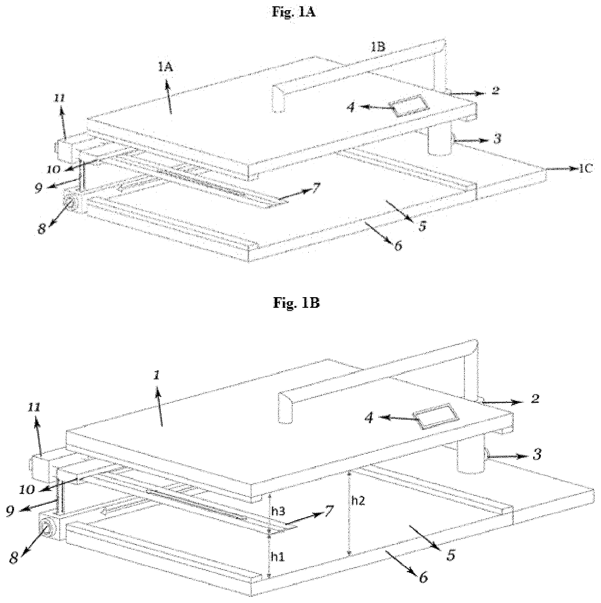
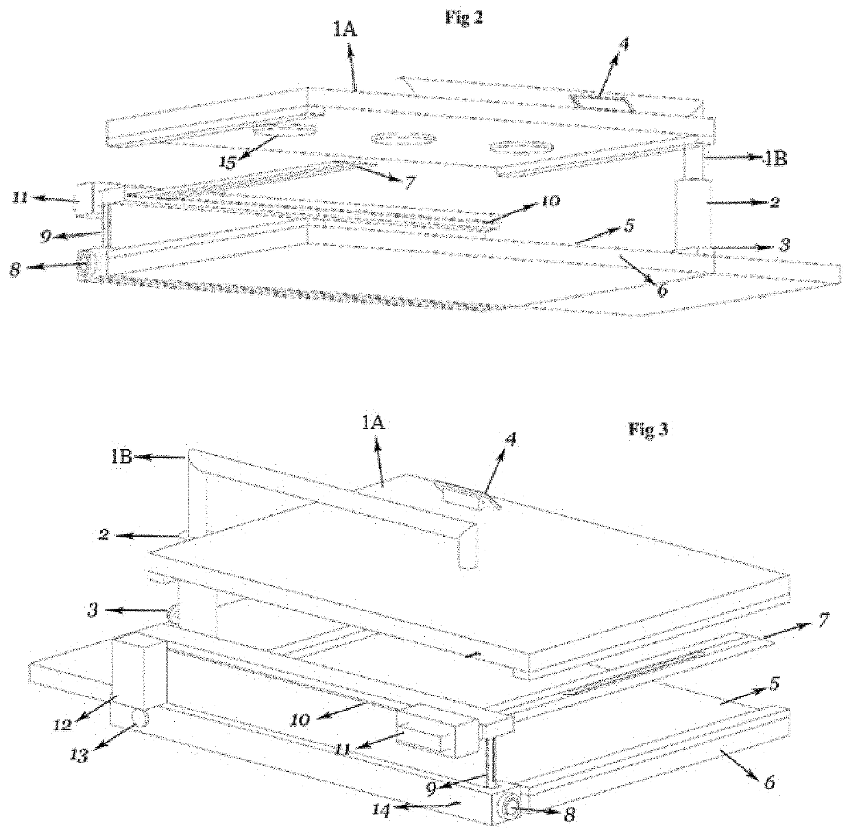
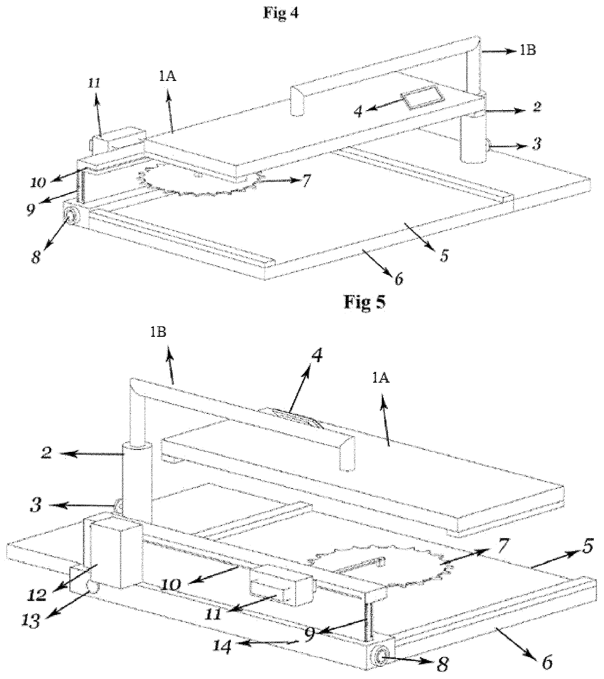
[0027]A tissue splitting device is disclosed which may be operated manually or automatically and which securely holds harvested tissue, especially soft tissue such as gingival tissue, during cutting so as to reproducibly provide layers of tissue having substantially uniform thicknesses. This tissue slices or layers produced by this device are viable, of substantially uniform thickness, and can be tailored using the device for particular surgical graft sites. Preferably the device and method work without requirement for embedment of the tissue during cutting and preferably provide adjustable sample thickness.

[0028]Embodiments of this technology include, but are not limited to the following.

[0029]One embodiment of the invention is a tissue splitting device comprising a clamp comprising a horizontal and parallel upper plate and lower mounting plate, and a horizontal space between the upper plate and lower mounting plate that can accommodate a harvested tissue, wherein a blade is horizo...


PUM


Abstract

A tissue splitting device for splitting a harvested soft tissue to provide a layer of tissue having a uniform thickness and methods for tissue transplantation using uniform layers of tissue.

Description

BACKGROUND OF THE INVENTIONField of Invention[0001]The present invention relates to the field of reconstructive surgery and particularly to a tissue splitting device that produces viable, undamaged and uniformly sized soft tissue layers for transplantation.Description of Related Art[0002]Soft tissue grafts are commonly used in reconstructive surgery including for gingival, dermal, and cartilage grafting and sometimes for bone grafting. Traditionally a surgeon manually trims a harvested tissue to prepare a graft having a desired size and thickness for implantation at a grafting site; Rijhwani et al., Free Gingival Autograft and Subepithelial Connective Tissue Graft for the Treatment of Gingival Recession: A Brief Review and Report of Three Cases. Seema Yadav Journal of Contemporary Dentistry, September-December 2016; 6(3):225-232; Harris R J. Creeping attachment associated with the connective tissue with partial-thickness double pedicle graft. J. Periodontol. 1997 September; 68(9):89...

Claims


Application Information

Patent Timeline
IPC IPC(8): A61B17/322B26D3/28B26D7/04A61B17/32A61B17/00B26D1/00B26D1/06A61B17/14
CPCA61B17/322B26D3/28B26D7/04A61B17/147A61B2017/0015A61B2017/00075A61B2017/00761A61B2017/00792A61B2017/00969A61B2017/3225A61B2017/320064B26D1/065B26D2001/004A61C8/0006
Inventor ZAHID, TALAL MANSOOR H.ALKHATEEB, ABDULHAMEED F.NADERSHAH, MOHAMMEDKHAN, EMAD ALINATTO, ZUHAIR SALEHALTTAS, SANA GHAZI
Owner KING ABDULAZIZ UNIV
Learn More