Controlled excitation and saturation of magnetisation transfer systems

a magnetisation transfer and excitation system technology, applied in the field of magnetic resonance imaging, can solve the problems of inability to clinically practicable, m0, t1 and t2 estimation errors, and undermine the utility of secure diagnostic methods, so as to achieve stable relaxation times, maintain control over the induced saturation of the background pool, and constant magnetisation transfer

Active Publication Date: 2021-11-30
KINGS COLLEGE LONDON
View PDF17 Cites 0 Cited by
  • Summary
  • Abstract
  • Description
  • Claims
  • Application Information

AI Technical Summary

Benefits of technology

[0008]Embodiments of the present invention relate to a system and associate method of MRI and MR spectroscopy which provide stable measurements of the relaxation times, T1 and T2, by using tailored multi-band RF pulses that allow direct control of the saturation conditions in the background pool of macro-molecular protons, and hence provide a flexible means to induce constant Magnetisation Transfer (MT) effects, or to achieve precisely varying MT effects. In the case of constant MR conditions, equal saturation of the background pool is obtained for all measurements independent of the parameters that may be changed, for example, the rotation rate used to obtain a desired flip angle, that is, the degree of change in the magnetisation of the free pool of protons. To achieve this, a new RF pulse is defined where the RF pulse energy is balanced between the free and the background protons such that different flip angles can be achieved in the free pool whilst maintaining control over the induced saturation of the background pool. In some applications, this helps to ensure that the effects of the background pool can be kept constant throughout the examination, and can therefore be effectively ignored. A further way of achieving this is to control the timing between the RF pulses to obtain overall constant saturation. This way, the total RF energy of each pulse is allowed to vary with varying flip angle, but the timing of the pulses adjusted to compensate for the changes in energy such that a constant level of saturation is maintained overall.
[0009]Under constant saturation, the measured signals effectively behave as a single pool of magnetization. This provides a reproducible measurement of M0 and T1, as well as removing errors in the estimation of T2. Consequently, this method of Controlled Saturation Magnetisation Transfer (CSMT) imaging makes it possible to achieve rapid generation of reproducible quantitative T1 and T2 maps using any of the currently available high resolution 3D clinical protocols (4, 10, 11, 18). In other applications CSMT imaging may be used to obtain a sequence of images each of which has precisely controlled MT conditions suitable, for example, for calculating the properties of both the free pool and the background pool.
[0010]A first aspect of the present invention provides a method of calculating a multi-band radio frequency (RF) pulse for use in magnetic resonance (MR) imaging or MR spectroscopy, wherein the multi-band RF pulse comprises an on-resonance band and at least one off-resonance band, and wherein the method comprises defining a target degree of rotation in the magnetisation, M, of a free pool of protons in an object, and calculating the parameters of the on-resonance band and the at least one off-resonance band based on the target degree of rotation, wherein the calculated parameters of the on-resonance band the at least one off-resonance band are such that, when the multi-band RF pulse is used as a parameter of MR imaging or spectroscopy, a predetermined amount of saturation occurs in a bound pool of protons in the object. As such, the parameters of the multi-band RF pulse are calculated such that they achieve the target degree of rotation in the free pool of protons, whilst maintaining a certain level of saturation of the background pool of protons when the RF pulse is used as a parameter of an MR scan. The calculated pulse therefore enables control of the saturation of the background pool.
[0019]The generating an image may further comprise calculating a myelin water fraction from the plurality of MR signals. By using the plurality of multi-band RF pulses to achieve controlled MT conditions, the reliability of the myelin water fraction estimation can be improved. The myelin water fraction is commonly used for understanding neurological conditions where the myelin in the brain starts to disappear, for example, multiple sclerosis. It is also measured to try to study brain development, since myelin is generated in the brain as a baby develops into early childhood.
[0023]The processor may be further caused to calculate a plurality of multi-band RF pulses corresponding to a plurality of target degrees of rotation, wherein the predetermined amount of saturation is constant for the plurality of multi-band RF pulses such that, when used as a parameter of MR imaging or spectroscopy, the plurality of multi-band RF pulses achieve controlled Magnetisation Transfer conditions. In some arrangements, the plurality of multi-band RF pulses achieve constant Magnetisation Transfer conditions. In this respect, the saturation effect on the background pool may be exactly the same for each RF pulse, or at least similar enough that only very small differences in the Magnetisation Transfer effects are exhibited, to the extent that single-pool assumptions are still valid. In other arrangements, the multi-band RF pulse is calculated so as to accurately control the degree of rotation of the free pool and the degree of saturation of the background pool.

Problems solved by technology

Current clinical protocols for quantitative imaging generally fail to take MT correctly into account, so producing variable results that undermine their utility as secure diagnostic methods (6).
Quantitative MT protocols can more precisely characterise tissue, but require more data to be collected so are not regarded as clinically feasible (9).
MT effects directly alter the measured tissue values of M0 and T1 and, if not accounted for, these then lead to errors in M0, T1 and T2 estimation.
If not considered, background pool effects necessarily biases relaxometry measurements as an extra source (or pathway) of magnetization recovery is interfering with the measured signal.
To avoid this, multi-compartment models have been proposed which require more measurements to be sampled and therefore induce longer scanning times, so hindering clinical applicability.

Method used


Image

  • Controlled excitation and saturation of magnetisation transfer systems
  • Controlled excitation and saturation of magnetisation transfer systems
  • Controlled excitation and saturation of magnetisation transfer systems

Examples


Embodiment Construction

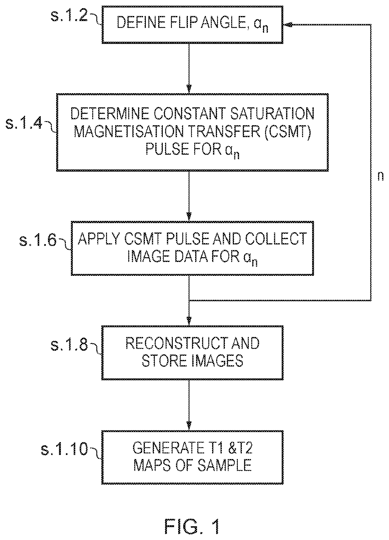
[0070]A brief overview of embodiments of the invention will now be given.

[0071]In biological samples, where multiple pools of magnetization are always present, applied RF pulses induce not only rotation of the free pool of protons but also a simultaneous partial saturation of the background pool of restricted protons. The background pool of protons is characterized by its short T2 (shorter than the duration of the RF pulse) and as a consequence does not produce an observable signal, however, since the energy of the applied RF pulses is absorbed by the background pool, this ultimately affects the relaxometry measurements of the free pool, specifically, the T1 measurement. This phenomenon is known as Magnetisation Transfer (MT). If the effects of MT are not taken into account, this can lead to errors in the T1 and T2 measurements. To take the saturation of the background pool into consideration, multi-pool modelling is usually required which is considerably time consuming. The present...


PUM

PropertyMeasurementUnit
degree of rotationaaaaaaaaaa
magnetisationaaaaaaaaaa
spectroscopyaaaaaaaaaa

Abstract

The present invention relate to a system and associate method of MRI and MR spectroscopy which provide stable measurements of the relaxation times, T1 and T2, by using tailored multi-band RF pulses that direct control of the saturation conditions in the background pool of macro-molecular protons, and hence provide a flexible means to induce constant Magnetisation Transfer (MT) effects. In doing this, equal saturation of the background pool is obtained for all measurements independent of the parameters that may be changed, for example, the rotation rate used to obtain a desired flip angle, that is, the degree of change in the magnetisation of the free pool of protons.

Description

TECHNICAL FIELD[0001]The present invention relates to a method and system for magnetic resonance (MR) imaging, and in some embodiments a method and system for MR imaging using tailored multi-band RF pulses to achieve controlled saturation of systems that exhibit Magnetisation Transfer (MT).BACKGROUND TO THE INVENTION AND PRIOR ART[0002]Unlike CT and PET, which both can provide images expressed in absolute units, Magnetic Resonance Imaging (MRI) is usually deployed as a highly sensitive qualitative modality. Thus, although conventional MRI is a first line investigation for both radiological diagnosis and treatment monitoring of neurological disease, almost all assessments are based on images presented in arbitrary units. There is growing interest in quantitative MRI as a potential route to less subjective diagnosis (1) and to allow cross site comparison such as for clinical trials. Key MR parameters are the proton density (M0) and the two relaxation times T1 and T2, which describe ho...

Claims


Application Information

Patent Timeline
Patent Type & Authority Patents(United States)
IPC IPC(8): G01V3/00G01R33/50G01R33/483G01R33/56G01R33/561
CPCG01R33/50G01R33/4838G01R33/5605G01R33/5614G01R33/5613G01R33/4616
Inventor TEIXEIRA, RUI PEDRO AZEREDO GOMESHAJNAL, JOSEPH VILMOSMALIK, SHAIHAN JALALWEST, DANIEL JOHN
Owner KINGS COLLEGE LONDON
Why Eureka
Try Eureka