Spect gamma camera

a gamma camera and spectrometer technology, applied in tomography, instruments, applications, etc., can solve the problems of reducing the sensitivity of the collimator, reducing the detection efficiency, and prolonging the data acquisition time, so as to improve the detection efficiency, improve the image resolution, and improve the detection efficiency.

Inactive Publication Date: 2003-11-06
ULTRASPECT
View PDF7 Cites 61 Cited by
  • Summary
  • Abstract
  • Description
  • Claims
  • Application Information

AI Technical Summary

Benefits of technology

0013] The technique of the present invention may lead to substantial improvement in image resolution (better than 7 mm) and improvement in image sensitivity with respect to the conve

Problems solved by technology

The sensitivity, however, is inversely related to geometric resolution, which means that improving collimator resolution decreases collimator sensitivity, and vice versa.
The current use of collim

Method used


Image

  • Spect gamma camera
  • Spect gamma camera
  • Spect gamma camera

Examples


Embodiment Construction

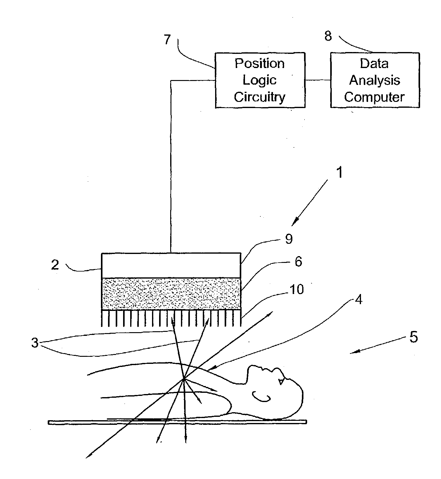
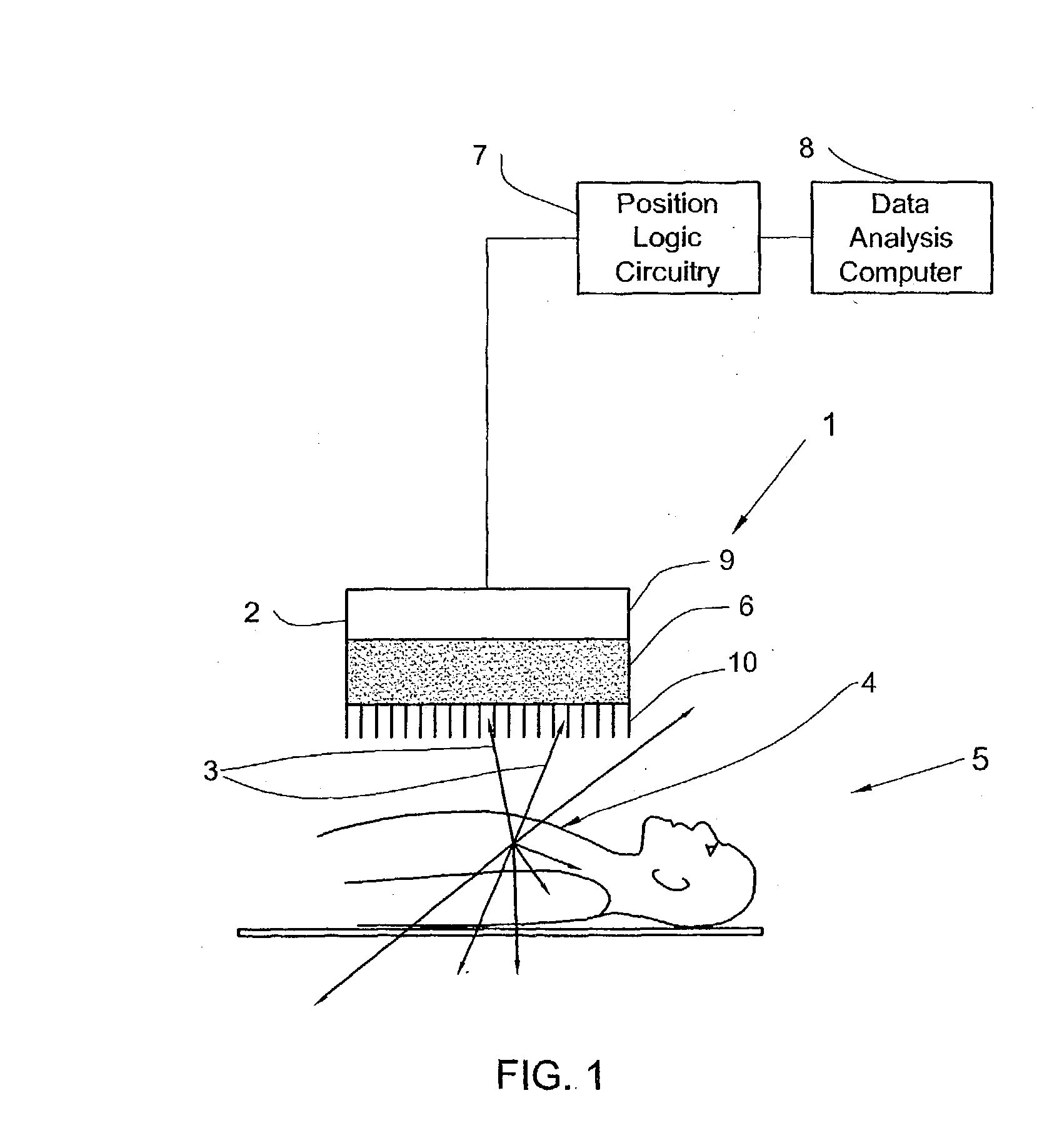
[0022] Reference first is made to FIG. 1 depicting a side view of a simplified schematic diagram of gamma camera in accordance with the present invention, for obtaining a SPECT image of a portion of a body that has been administered by a radiopharmaceutical substance which radiates gamma rays.

[0023] The gamma camera 1 comprises a detector 2 mounted above an inspected portion 4 of a body 5, a position logic circuitry 7 and a data analysis computer 8, all connected appropriately.

[0024] Detector 2 includes at least one photon detector crystal 6 facing the portion 4 of body 5. The photon detector crystal 6 may be in the form of a semiconductor crystal or crystals. This crystal(s) may be selected from a first group including Cadmium-Telluride (CdTe), Cadmium-Zinc-Telluride (CeZnTe), Lead Iodine (PbI).

[0025] The detector 2 of the gamma camera 1 may further include at least one photo-multiplier 9. The photon detector crystal(s) in this case may be selected from a second group including Sod...


PUM


Abstract

A method and apparatus of obtaining and reconstructing an image of a portion of a body, administered by a radiopharmaceutical substance, by using Single-photon emission computerized tomography (SPECT) for determination of functional information thereon. The method comprises (a) acquiring gamma ray photons emitted from said portion by means of a detector capable of converting the photons into electric signals, the detector having at least one crystal and allowing said gamma rays having incident angles essentially exceeding 5 degrees and, preferably, exceeding 10 degrees to be detected; (b) processing said electric signals by a position logic circuitry and thereby transforming them into data indicative of positions on said photon detector crystal, where the photons have impinged the detector; and (c) reconstructing an image of a spatial distribution of the pharmaceutical substance within the portion of the body by processing said data and taking into consideration weight values which are functions of angles and, possibly, distances between different elements of the portion of the body and corresponding elements of this position's projection on the detector.

Description

[0001] This invention relates to Nuclear Medicine, and, more particularly, to Single-photon emission computerized tomography (SPECT) imaging technique.[0002] SPECT is one of Nuclear Medicine imaging techniques that enable to acquire functional information about patient's specific organ or body system. This functional information is attained from analysis of internal radiation obtained from pharmaceutical substance administered to the patient, which is labeled with a radioactive isotope. The radioactive isotope decays, resulting in the emission of gamma rays, thus providing with information on the concentration of the radiopharmaceutical substance introduced to the patient's body. An instrument for the detection of gamma ray emissions of the radiopharmaceutical substance administered in the body is known as gamma camera. The SPECT technique collects gamma ray photons that are emitted from the patient and reconstructs an image or a series of images of the place in the body from which ...

Claims


Application Information

Patent Timeline
IPC IPC(8): A61B5/05G01TG01T1/164G01T1/172
CPCA61B6/037G01T1/1644
Inventor SHWARTZ, SHOULAMIT C.OHANA, ISRAEL
Owner ULTRASPECT
Why Eureka
Try Eureka