Ultrasonic microkeratome

a microkeratome and ultrasonic technology, applied in the field of ultrasonic microkeratome, can solve the problems of low error margin, complex procedure and instrumentation, and inability to perform surgery with a single device,

Inactive Publication Date: 2004-01-01
SLADE STEPHEN G
View PDF43 Cites 10 Cited by
  • Summary
  • Abstract
  • Description
  • Claims
  • Application Information

AI Technical Summary

Problems solved by technology

Due to poor success with removing stroma from the bed either manually with the Paufique knife or a manual keratome he abandoned the in situ approach and refined the lamellar dissection and precise carving of the corneal lamellar disc.
Several drawbacks, however, were inherent to Dr.
Barraquer's initial techniques which included the complex nature of the procedure and instrumentation, low margin for error, and steep surgeon learning curve.
After the laser application, the epithelium must regrow which can be a painful process with reduced vision for several days.
Further, the development of postoperative scarring in the central cornea after excimer laser surface ablations has resulted in regression of effect, significant disturbing visual complaints and lines of lost best-corrected vision.
Additionally, the postoperative discomfort and the relatively long postoperative recuperative period after surface ablation is currently an inescapable reality for both the patient and the eye care professional.
While LASIK offers several of advantages over PRK, the creation of the corneal flap has been associated with a number of intra-operative and post-operative complications.
Thus some patients with high myopia or thin corneas are left without a treatment option.
Second, cutting the flap in the cornea alters the biomechanical properties of the cornea and may induce aberrations or irregularities in the surface of the cornea.
Third, the incidence of complications involving the flap, while small, is significant.
Also, to help remove the epithelium drugs such as alcohol must be used in LASEK to loosen the epithelium, damaging the epithelium and slowing recovery.
As a result, patients have more discomfort and a slower return of good vision.
The technique also is surgically tedious and difficult.

Method used


Image

  • Ultrasonic microkeratome
  • Ultrasonic microkeratome
  • Ultrasonic microkeratome

Examples


Embodiment Construction

[0035] The eye works on a principle very similar to that of a camera. The iris or colored portion of the eye about the pupil, functions like a shutter to regulate the amount of light admitted to the interior of the eye.

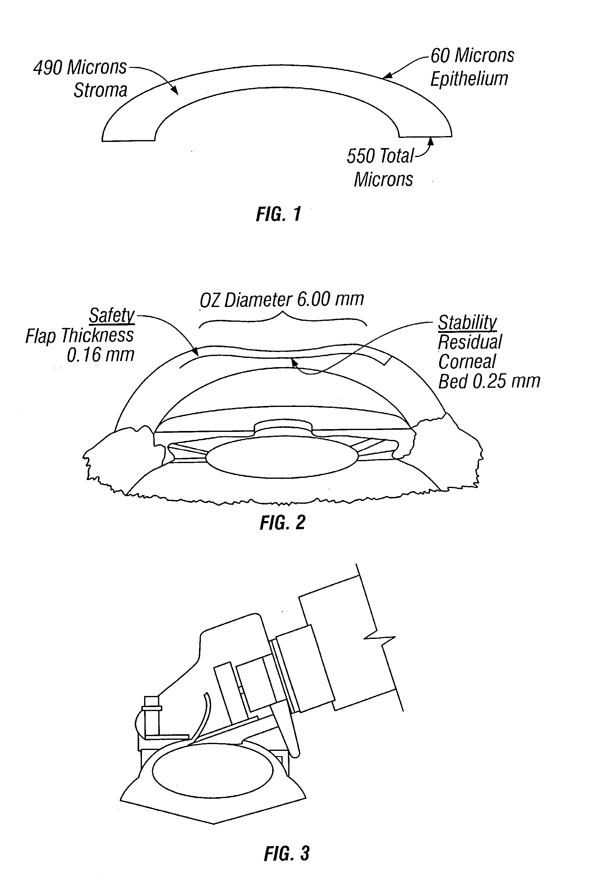
[0036] The cornea or clear window of the eye, and the lens, which is located behind the pupil, serve to focus the light rays from an object being viewed onto the retina at the back of the eye. The cornea is composed of five layers; first the epithelium that is five cells thick and is usually around 60 microns thick. A thin membrane called Bowman's membrane underlies the epithelium. The mass of the cornea is called the stroma, which is about 480 microns thick. The fourth layer is another, stronger but very thin membrane called Descemet's. The final layer is the endothelium, which is only one cell thick. Bowman's, Descemet's and the endothelium do not contribute significantly to the total cornea thickness. The total thickness of the cornea averages around 540 microns. O...


PUM


Abstract

The invention relates to medical instruments and methods for performing eye surgery to correct focusing deficiencies of the cornea. More particularly, the present invention relates to mechanical instruments known as microkeratomes, and related surgical methods for performing lamellar keratotomies and refractive surgery. The device is designed to create a flap of epithelium, or stroma and epithelial flap as is done now with LASIK. This will enable a new technique, ELF, or Epithelial Laser Flap, which will combine the advantages of LASIK and PRK, an older, but easier technique.

Description

[0001] This application claims priority to U.S. Provisional Application 60 / 362,305, filed Mar. 7, 2002.[0002] Not Applicable.REFERENCE TO A MICROFICHE APPENDIX[0003] Not Applicable.[0004] 1. Field of the Invention[0005] The present invention relates to medical instruments and methods for performing eye surgery to correct irregularities of the cornea. More particularly, the present invention relates to mechanical instruments known as microkeratomes, and related surgical methods for performing lamellar keratotomies and refractive surgery.[0006] 2. General Background of the Invention[0007] In recent years, as Refractive Surgery has developed, a number of surgical techniques have become available to surgically treat near sightedness, farsightedness and astigmatism. Of these surgical techniques, laser in situ keratomileusis (LASIK) has evolved into one of the most promising members in the family of lamellar refractive surgeries. LASIK has recently gained popularity for the correction of ...

Claims


Application Information

Patent Timeline
Patent Type & Authority Applications(United States)
IPC IPC(8): A61F9/013
CPCA61F9/0133A61F9/013
Inventor SLADE, STEPHEN G.
Owner SLADE STEPHEN G
Why Eureka
Try Eureka