Methods of detecting cancer cells in biological samples

a biological sample and cancer cell technology, applied in the field of methods of detecting cancer cells in biological samples, can solve the problems of rare cancer cells, highly curable grade i tumors are virtually undetectable using cytological staining and cytological staining methods

Inactive Publication Date: 2004-10-07
BIOVIEW
View PDF6 Cites 43 Cited by
  • Summary
  • Abstract
  • Description
  • Claims
  • Application Information

AI Technical Summary

Problems solved by technology

However, at lower grades of this progression, cancerous cells are rare and in some cases appear similar to those seen in other conditions not related to cancer such as inflammation, obstruction or stones.
However, the highly curable grade I tumors are virtually undetectable using cytological staining.
Cytological staining methods have several other disadvantages especially in cases where there are no identifiable tumors or pre-cancerous lesions.
In general, each of these markers has better sensitivity than cytology alone but is prone to more false-positive findings.
Thus, the current methods of assessing biological specimens are time consuming and are prone to diagnostic errors resulting from missing slide areas and misinterpretation of the stained sample.
In addition, since current approaches utilize a single staining method at a time, such approaches increase the chance of either false negative

Method used


Image

  • Methods of detecting cancer cells in biological samples
  • Methods of detecting cancer cells in biological samples
  • Methods of detecting cancer cells in biological samples

Examples


example 1

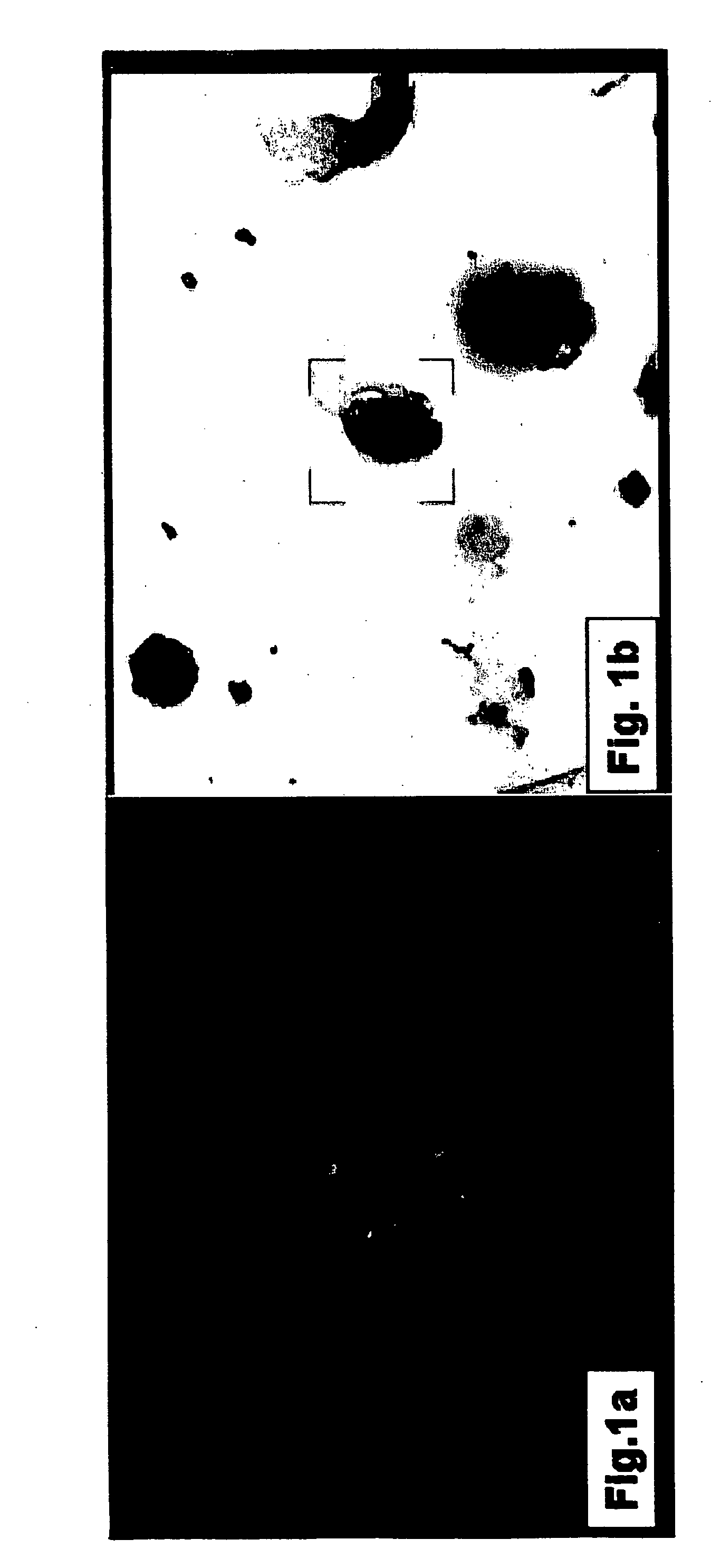
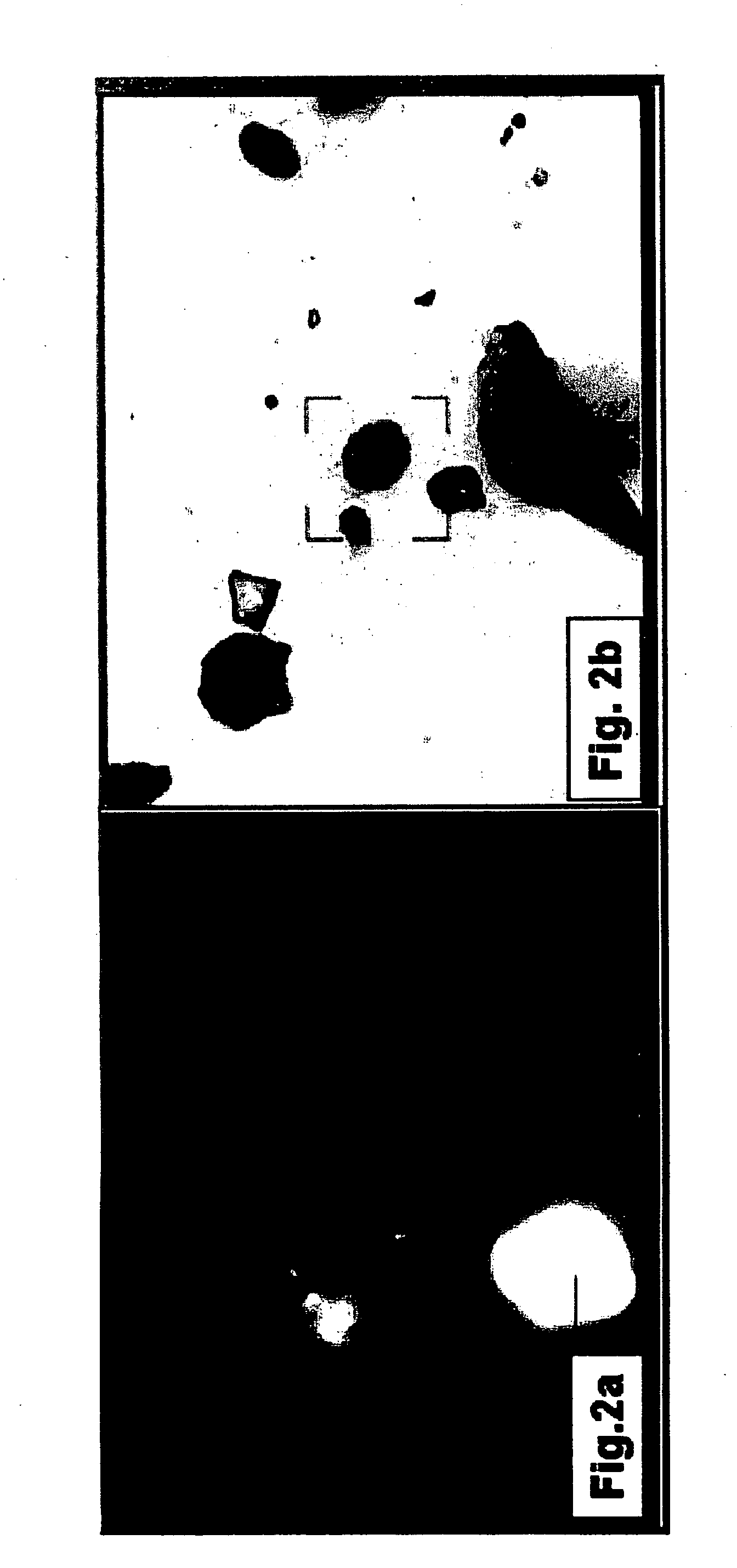
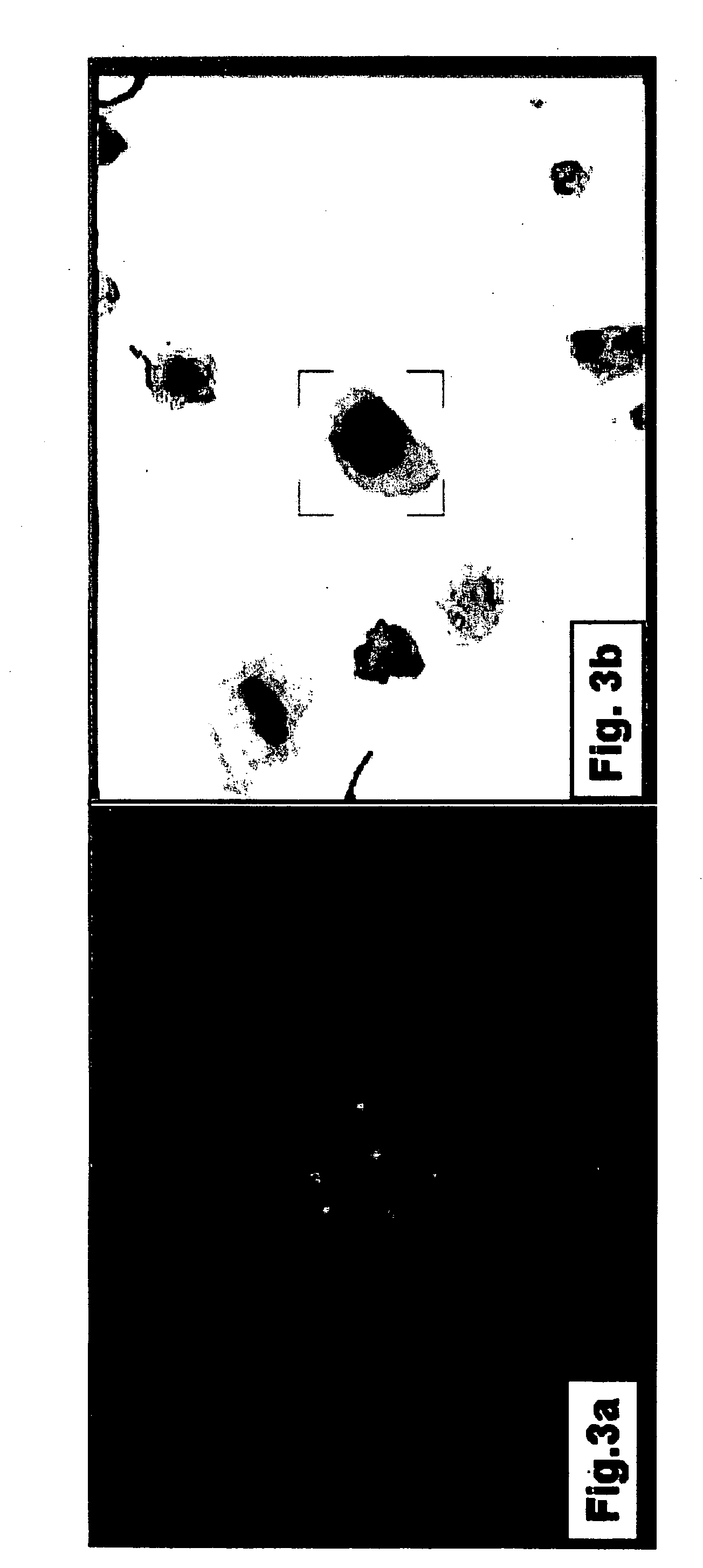
[0139] Transitional Cell Carcinoma is Accurately Detected Using Double Staining and Dual Imaging

[0140] In order to test the suitability of the combined staining / dual imaging method of the present invention in identifying transitional cell carcinoma (TCC), voided urine samples were subjected to morphology staining followed by FISH analysis and the staining results were analyzed using dual imaging.

[0141] Materials and Experimental Methods

[0142] Preparation of cytospin slides of voided urine samples--Voided urine samples (volume ranged from 4 ml to 45 ml, mean 15.+-.11.3 ml) were centrifuged at room temperature for 10 minutes at 300.times.g. Following centrifugation cell pellets were resuspended in 100-300 .mu.l of a Morphology Preserver (BioWhite kit, BioView LtD., Rehovot, Israel) and the concentration of cells was determined using a Neubauer improved counting chamber (Neubauer, Germany). Cells were then cytospun at a cell density of 300-500 cells per mm.sup.2 according to manufactur...

example 2

[0154] A Combined Dual Staining / Dual Imaging Method is Highly Sensitive in Diagnosing TCC

[0155] In order to test the sensitivity of the combined staining / dual imaging method of the present invention in diagnosing transitional cell carcinoma (TCC), the diagnostic scores obtained using this method were compared with those obtained by urine cytology, cystoscopy and bladder biopsies.

[0156] Experimental and Statistical Results

[0157] Comparative analysis of screening methods for transitional cell carcinoma (TCC)--Thirty five urine samples were screened for the presence of TCC using either a morphological stain alone (see "cytology" in Table 1, hereinbelow) or a combined morphological / FISH dual imaging method (see "combined" in Table 1, hereinbelow). The accuracy of TCC diagnosis was compared to concurrent cystoscopy findings and pathological examination of bladder biopsies.

1TABLE 1 Comparison of various detection methods for transitional cell carcinoma Mix I Mix II No. of No. of abnorm. a...


PUM


Abstract

The invention provides methods of detecting cancerous cells in biological samples using a double staining/dual imaging approach, which can be used to diagnose cancer. More specifically, the present invention provides methods of diagnosing bladder cancer by a simultaneous scanning of cell morphology and FISH signals of cells derived from a urine sample.

Description

[0001] This application claims the benefit of priority from U.S. provisional patent application No. 60 / 459,992, filed Apr. 4, 2003.FIELD AND BACKGROUND OF THE INVENTION[0002] The present invention relates to methods of detecting cancer cells in biological samples using a double staining / dual imaging approach, more particularly, embodiments of the present invention relate to a method of detecting transitional cell carcinoma in voided urine samples using dual imaging with consecutive scanning of cell morphology and FISH signals.[0003] Early detection of cancer is the key feature in treating cancer patients. For many types of cancer, such as breast cancer, detection is often possible via physical examination of the cancer tissue. In other types of cancer, such as in leukemia, cancer detection is based on the examination of cancerous cells in blood or bone marrow samples, while in kidney or bladder cancers, the cancerous cells can be detected in voided urine. Thus the identification of ...

Claims


Application Information

Patent Timeline
IPC IPC(8): G01N1/30G01N33/574
CPCG01N1/30G01N33/57484
Inventor DANIELY, MICHALKAPLAN, TALKAPLAN, ERANFREIBERGER, AVNER
Owner BIOVIEW
Why Eureka
Try Eureka