Autofluorescence detection and imaging of bladder cancer realized through a cystoscope

a cystoscope and autofluorescence technology, applied in the direction of fluorescence/phosphorescence, spectroscopy, instruments, etc., can solve the problems of short photon penetration depth in tissues, time-consuming and bulky diagnostic equipment, and extraction of information, etc., to achieve cost-effective effects

Inactive Publication Date: 2008-02-28
LAWRENCE LIVERMORE NAT SECURITY LLC
View PDF11 Cites 25 Cited by
  • Summary
  • Abstract
  • Description
  • Claims
  • Application Information

AI Technical Summary

Benefits of technology

[0008] Accordingly, the present invention provides a diagnostic apparatus that includes: at least one electromagnetic radiation source, an arranged interior examination device adapted to transmit the radiation source and further adapted to relay a near-infrared scattered and / or an autofluorescence emission radiation, and a detector adapted to capture the scattered and / or near-infrared emission radiation produced by the radiation sources and optically coupled to the interior examination device so that characterization of the one or more tissue components is capable of being performed.
[0009] Another aspect of the present invention provides a diagnostic apparatus that includes: at least two substantially narrow-band electromagnetic radiation wavelength sources for simultaneous illumination of one or more tissue components, an arranged interior examination device adapted to transmit the wavelength sources for illumination of the tissue components and further adapted to relay a near-infrared scattered radiation of the illuminated tissue components; and a detector adapted to simultaneously capture one or more images produced by the near-infrared scattered radiation from the tissue components so that characterization of the one or more tissue components is capable of being performed.
[0013] Accordingly, the present tissue imaging system and method provides a portable, cost effective, non-invasive arrangement, capable of directing substantially monochromatic electromagnetic radiation and capable of providing spectrally processed images in the much desired need for differentiating components in human and / or animal tissues.

Problems solved by technology

Diagnostic medical equipment typically includes time-consuming, bulky, expensive apparatus that often exposes human tissue components to potentially harmful radiation and or chemicals.
The disadvantage of these wavelengths is their short photon penetration depth in tissues, which leads to extraction of information only from superficial tissue layers.

Method used


Image

  • Autofluorescence detection and imaging of bladder cancer realized through a cystoscope
  • Autofluorescence detection and imaging of bladder cancer realized through a cystoscope
  • Autofluorescence detection and imaging of bladder cancer realized through a cystoscope

Examples


Embodiment Construction

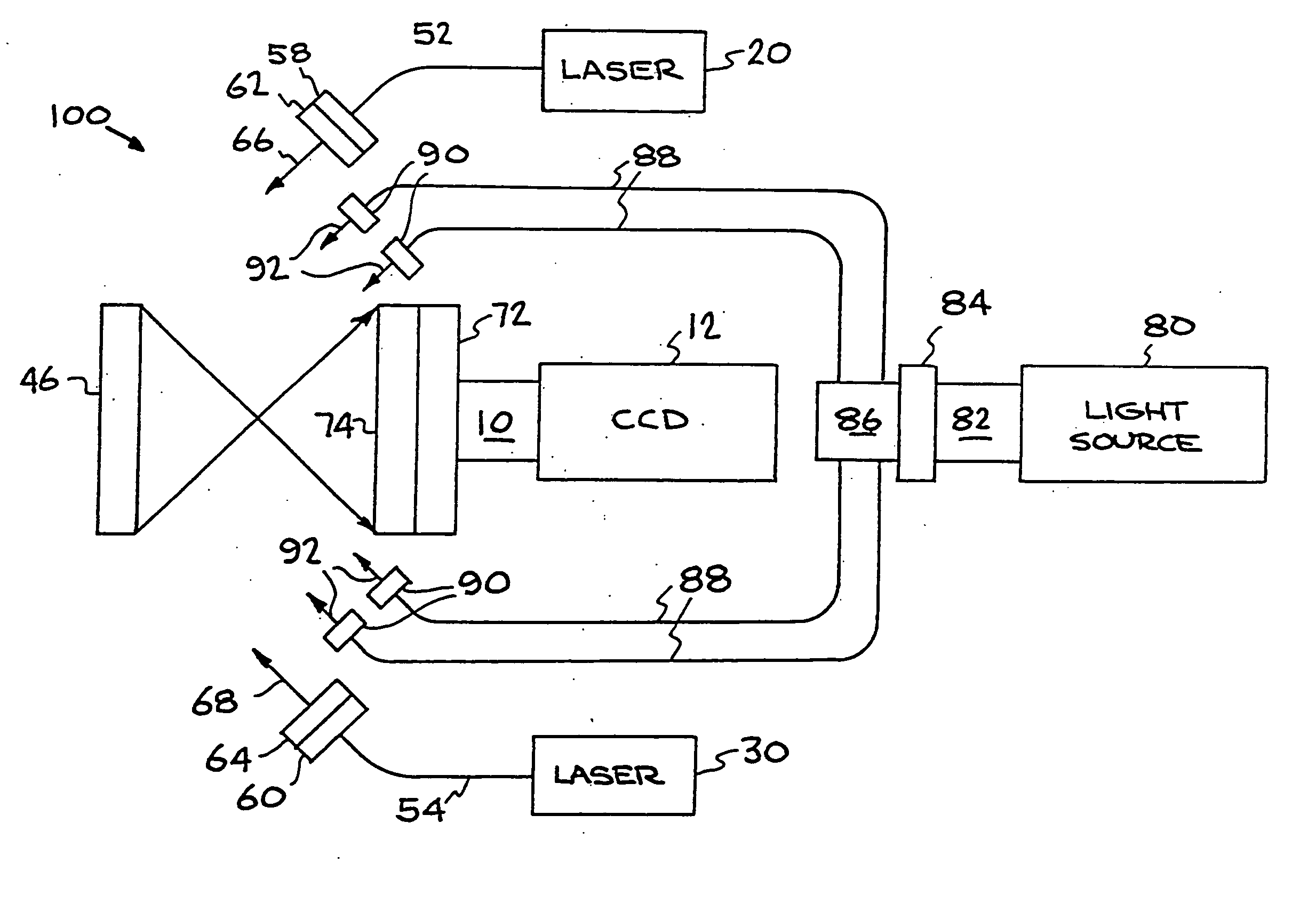
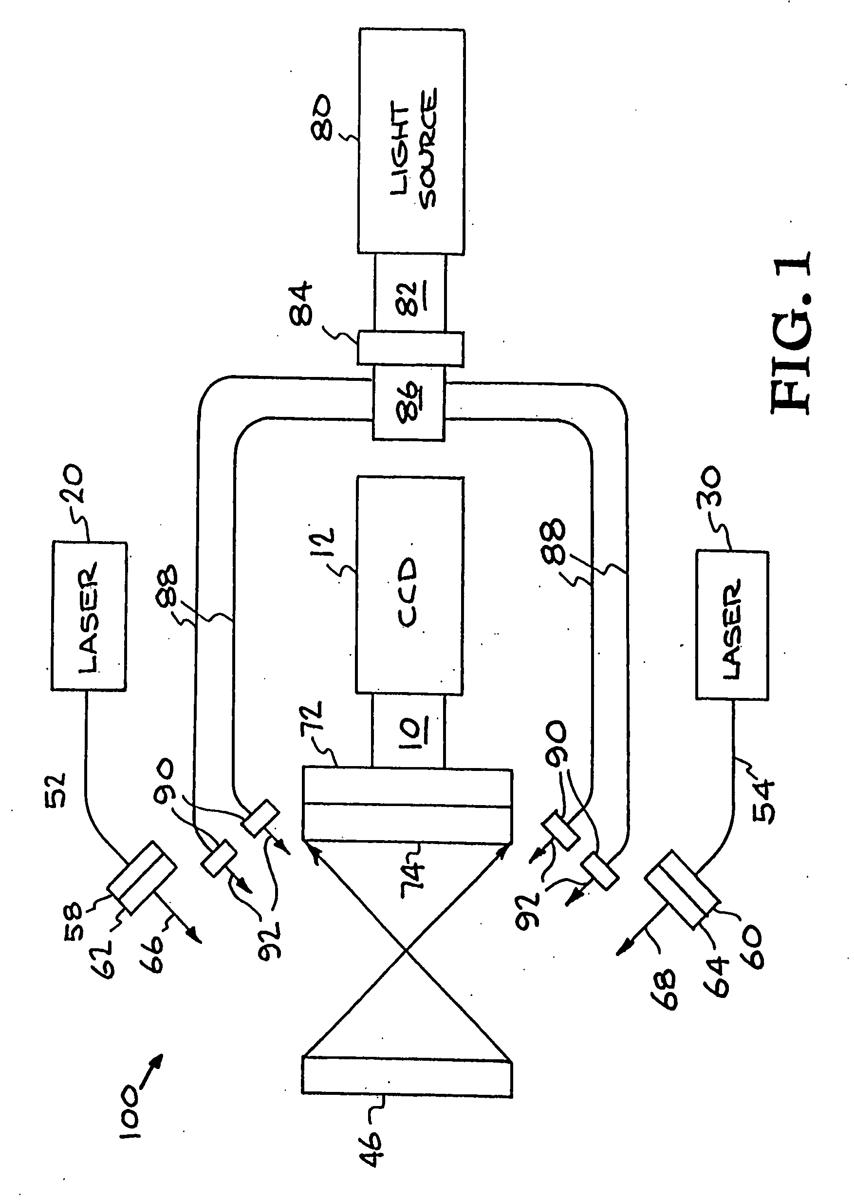
[0022] Referring now to the drawings, specific embodiments of the invention are shown. The detailed description of the specific embodiments, together with the general description of the invention, serves to explain the principles of the invention.

[0023] Unless otherwise indicated, all numbers expressing quantities of ingredients, constituents, reaction conditions and so forth used in the specification and claims are to be understood as being modified in all instances by the term “about.” Accordingly, unless indicated to the contrary, the numerical parameters set forth in the specification and attached claims are approximations that may vary depending upon the desired properties sought to be obtained by the subject matter presented herein. At the very least, and not as an attempt to limit the application of the doctrine of equivalents to the scope of the claims, each numerical parameter should at least be construed in light of the number of reported significant digits and by applyin...


PUM

PropertyMeasurementUnit
wavelengthsaaaaaaaaaa
wavelengthaaaaaaaaaa
output poweraaaaaaaaaa

Abstract

Near infrared imaging using elastic light scattering and tissue autofluorescence and utilizing interior examination techniques and equipment are explored for medical applications. The approach involves imaging using cross-polarized elastic light scattering and / or tissue autofluorescence in the Near Infra-Red (NIR) coupled with image processing and inter-image operations to differentiate human tissue components.

Description

RELATED APPLICATION [0001] This application is a division of U.S. application Ser. No. 10 / 400,024, filed Mar. 25, 2003, entitled “Autofluorescence Detection and Imaging of Bladder Cancer Realized Through a Cystoscope,” which is a Continuation-In-Part of application Ser. No. 10 / 190,231 filed Jul. 5, 2002, and claims priority thereto.[0002] The United States Government has rights in this invention pursuant to Contract No. W-7405-ENG-48 between the United States Department of Energy and the University of California for the operation of Lawrence Livermore National Laboratory.BACKGROUND OF THE INVENTION [0003] 1. Field of the Invention [0004] The present invention relates to a medical diagnostic for the examination of tissue components. Specifically, the present invention relates to an optical imaging method and apparatus for in-vivo and real-time imaging of bladder cancer and determination of tumor margins. [0005] 2. Description of Related Art [0006] Diagnostic medical equipment typical...

Claims


Application Information

Patent Timeline
Patent Type & Authority Applications(United States)
IPC IPC(8): A61B6/00G01N21/49G01N21/64
CPCA61B1/043A61B1/0638A61B1/307A61B1/313A61B5/0071A61B5/0075A61B1/0669G01N21/49G01N21/6445A61B1/00186A61B1/063A61B1/0646A61B5/0086
Inventor DEMOS, STAVROS G.WHITE, RALPH W. DEVERE
Owner LAWRENCE LIVERMORE NAT SECURITY LLC
Why Eureka
Try Eureka