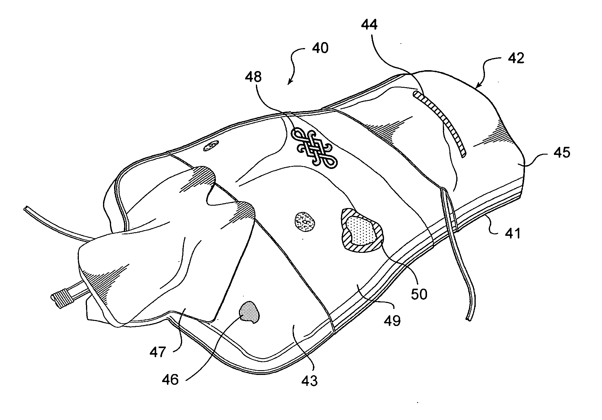
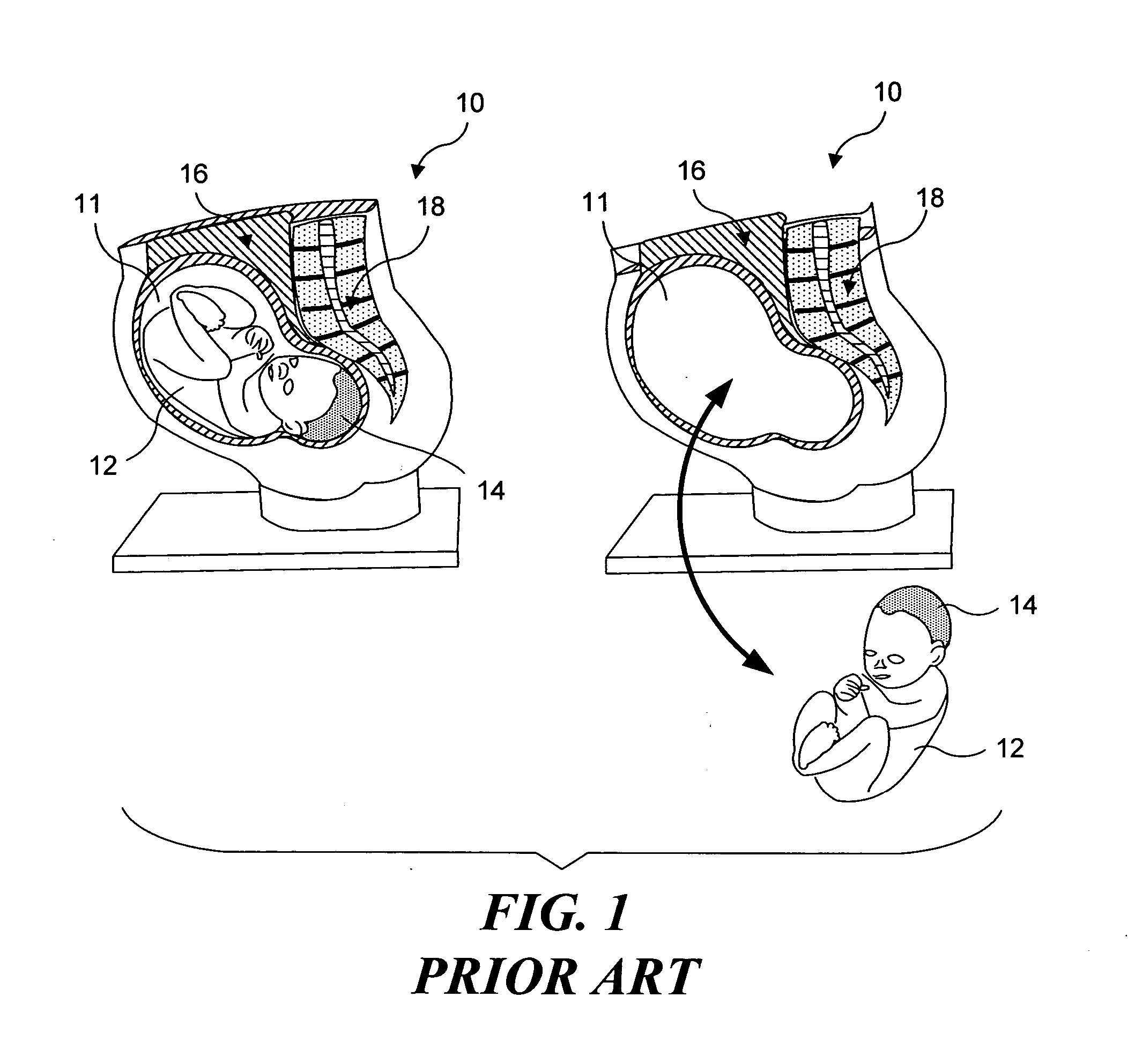
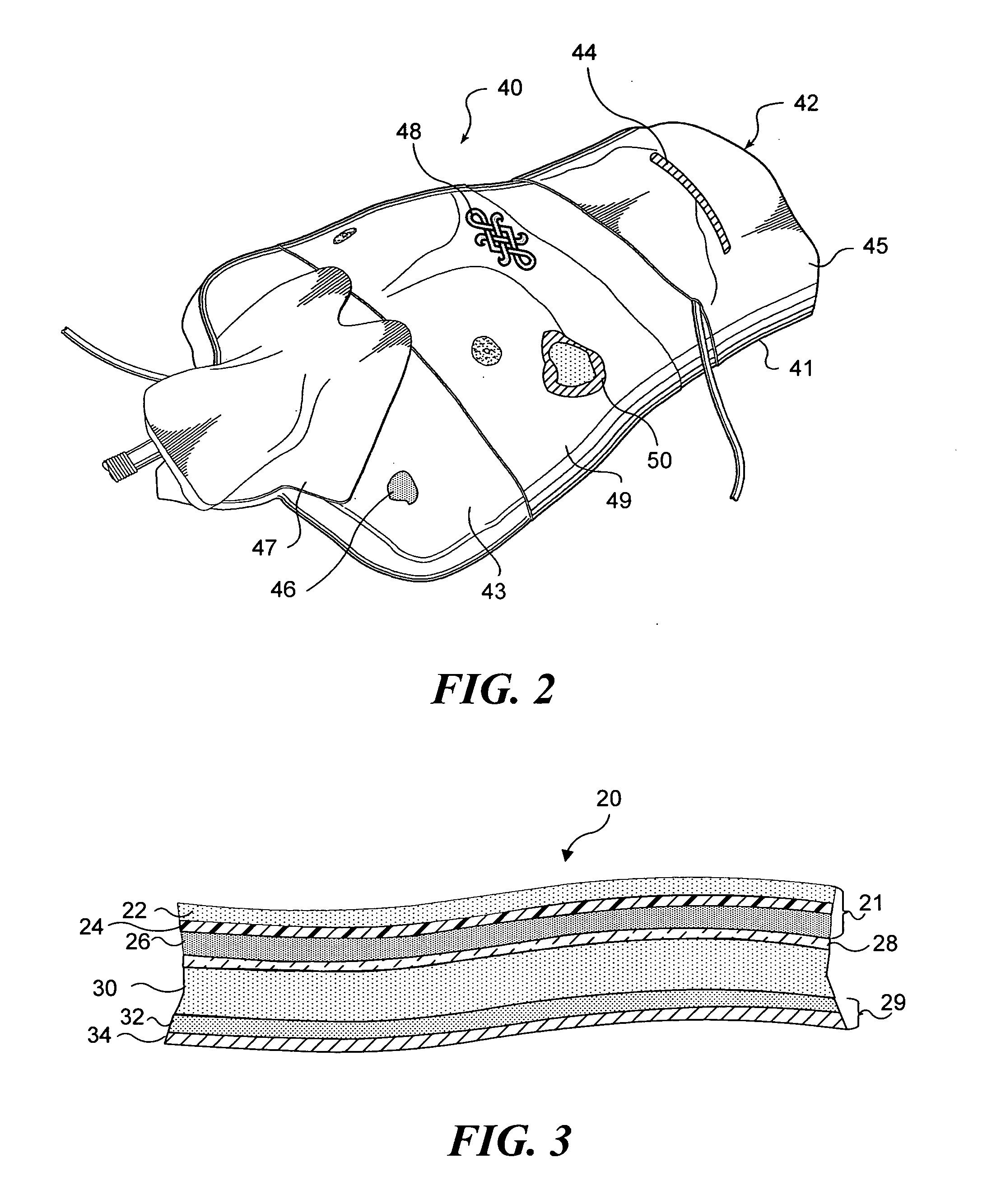
[0010] A single simulated physiological structure can include a plurality of different image
layers. For example, a simulated physiological structure representing a simulated tissue structure (and having dermal and sub-dermal tissue
layers) includes a plurality of different layers of elastomeric materials. A plurality of image layers are preferably incorporated into such a simulated physiological structure to enhance the realism of the simulated physiological structure. A first image layer corresponds to
skin, and includes variations in shading and color, as exhibited by real
human skin. Preferably, the image layer will be printed onto a fabric substrate, using a
real image of
human skin as the source of the image. Such an image should include features normally associated with
human skin, including such features as hair, freckles, variations in color and shading, and occasional imperfections, such as
scars, bruises, abrasions, and tattoos. The resulting image layer is coupled to an elastomeric material selected to represent a dermal layer. If desired, a
thin layer of relatively clear elastomeric material can be placed over the image layer, so that the upper surface of the simulated tissue structure has the tactile feel of tissue and does not look or feel like fabric. An image layer can also optionally include
skin abnormalities such as wounds.
[0012] The
specific substrate selected for use in an image layer will be a function of the specific simulated physiological structure, and a disposition of the image layer with respect to the specific simulated physiological structure. In a simulated tissue structure, where such a simulated tissue structure is configured to be incisable, adding fibrous layers (having different thicknesses and textures) at selected positions can enhance the realism of the simulated tissue structure so that the simulated tissue structure responds to incisions much like real tissue. A
fibrous layer can be included adjacent to the dermal layer, so that the dermal layer will provide more resistance to incision than a sub-dermal layer, which corresponds to softer tissue. Additional fibrous layers can be added deeper into the simulated tissue structure, to simulate serous membranes. Where an image layer is desired adjacent to a
fibrous layer that is included to enhance a tactile response (for example, near the dermal layer), the substrate onto which the image is to be printed can be the
fibrous layer selected to enhance the tactile realism of the simulated physiological structure. Also, where an image layer is desired, and no fibrous layer is needed for tactile response (or where inclusion of a fibrous layer would provide an unrealistic tactile response), a substantially non-fibrous substrate can be selected. For example, a plastic substrate can be used in place of a fabric or fibrous substrate. Presentations using overhead projectors often employ images and text that have been printed onto transparent plastic sheets (generally referred to as transparency films). Such transparent sheets can be used as a substrate material where a fibrous substrate is undesirable.
[0013] Selecting an image of tissue exhibiting a diseased or abnormal condition, for incorporating into an image layer enables very realistic training models to be achieved. For example, an image of a wound that penetrates the
skin layer can be selected for an image layer. The image layer is then incorporated into a simulated tissue structure to achieve a simulated tissue structure that accurately visual depicts a wound. Because wounds often change the contours of the skin layers at the
wound site, the contours of the wound can be reproduced in an elastomeric layer corresponding to a dermal tissue layer as it would appear if affected by an actual wound. The image layer is draped over the contoured elastomeric layer, so that the image of the wound is properly positioned relative to the contours of the elastomeric layer.
[0014] Images of
disease conditions associated with organs can also be employed. For example, a simulated
stomach might have an elastomeric layer simulating the lining of the
stomach. An image of a
stomach ulcer can be selected, and an image layer generated using this image. The resulting image layer is coupled to the elastomeric layer to achieve a realistically appearing simulated physiological structure (a model of a stomach including an ulcer). As noted above, if desired, the elastomeric layer simulating the stomach lining can include contours corresponding to those of a true ulcer. When the image layer is properly positioned relative to the elastomeric layer, the model can be used for video endoscopic training exercises. A student examining this model with a real or simulated
endoscope can examine the interior of the stomach and discover a visually realistic appearing ulcer.

