

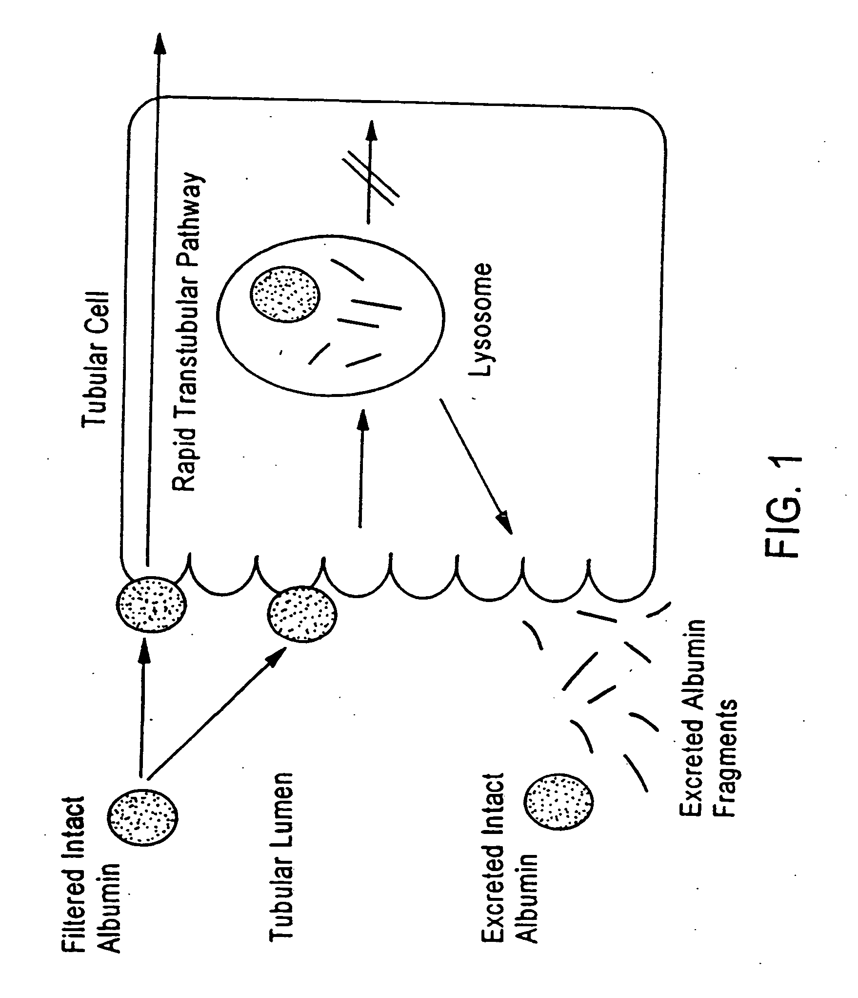
Method for kidney disease detection
a technology for kidney disease and detection methods, applied in the field of kidney disease detection, can solve the problem that filtering proteins currently does not detect the intact form of these proteins
- Summary
- Abstract
- Description
- Claims
- Application Information
AI Technical Summary
Benefits of technology
Problems solved by technology
Method used
Image

Examples
example 1
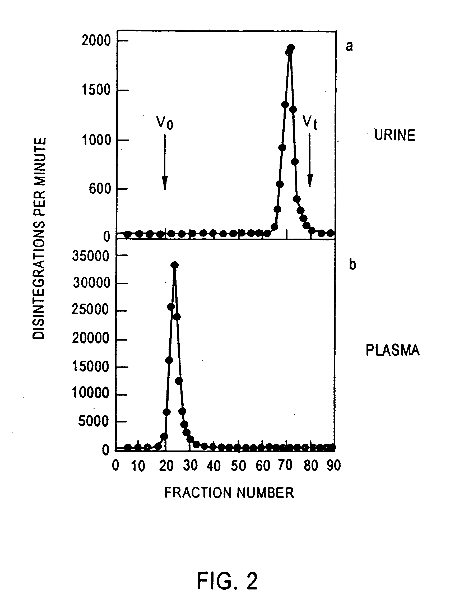
Size Exclusion Chromatography of Human Serum Albumin (HSA)
Normal, healthy volunteers were used to provide urine for analyzing the distribution of albumin in their urine.
3H[HSA] (Human Serum Albumin) was injected into healthy volunteers and urine and plasma were collected and analyzed by size exclusion chromatography using a G-100 column. The column was eluted with PBS (pH=7.4) at 20 ml / hr at 4° C. The void volume (V0) of the column was determined with blue dextran T2000 and the total volume with tritiated water.
Tritium radioactivity was determined in 1 ml aqueous samples with 3 ml scintillant and measured on a Wallac 1410 liquid scintillation counter (Wallac Turku, Finland).
FIG. 2 illustrates the distribution of albumin in urine and in plasma.
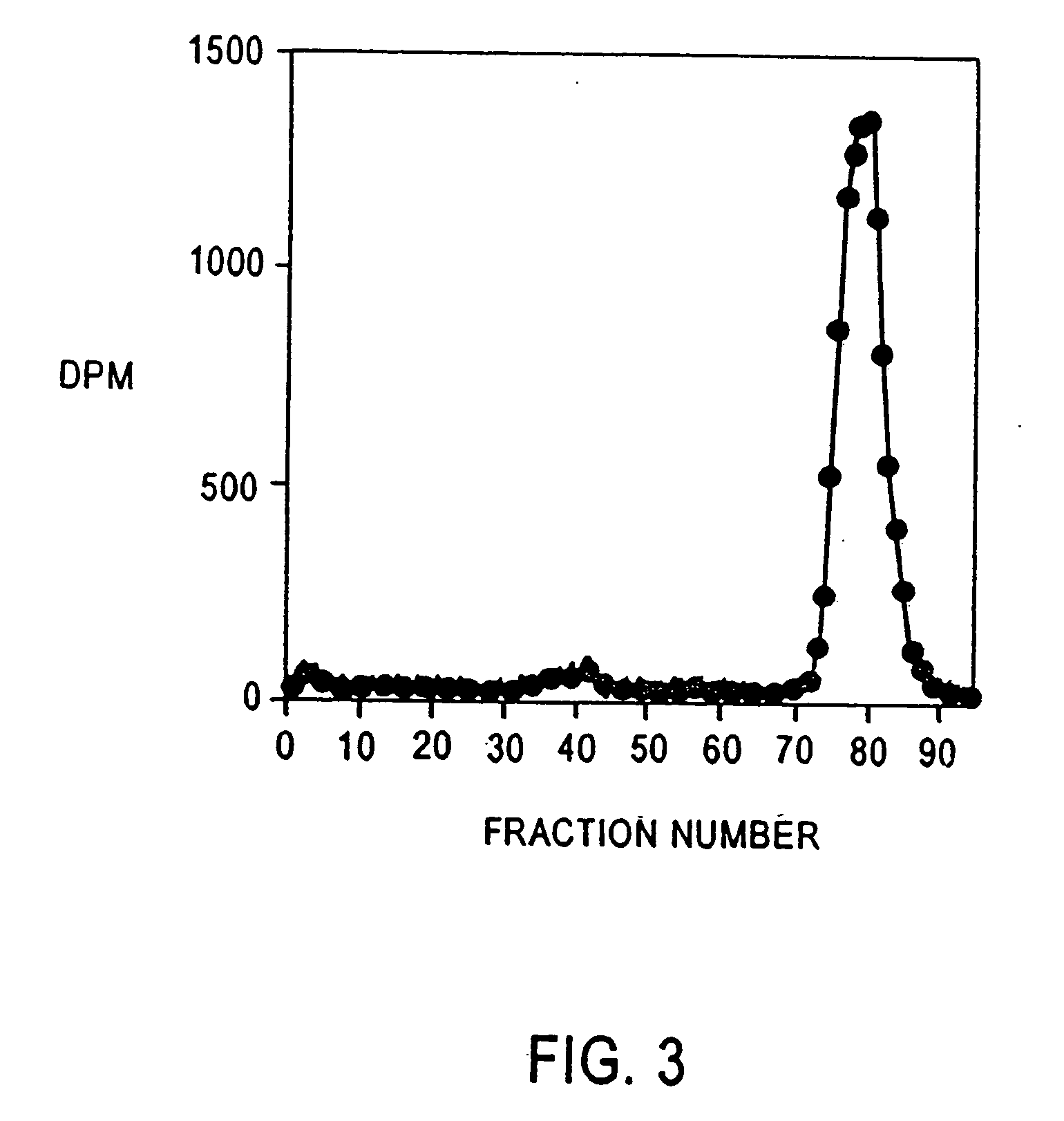
example 2
Albumin Excretion in a Normal, Healthy Volunteer and Diabetic Patient 3H[HSA] as used in Example I was injected into a normal, healthy volunteer and a diabetic patient. Samples of urine were collected and 3H[HSA] was determined as in Example 1.
The normal, healthy volunteer (FIG. 3) shows the excretion of fragments of albumin on a size exclusion chromatography as performed in Example 1.
The diabetic patient (FIG. 4) shows the presence of substantially full-length and fragmented albumin on size exclusion chromatography. However, excretion rates of albumin detectable by these methods were in the order of 5 μg / min (control) and 1457 μg / min (diabetic).
example 3
Determination of Intact Albumin, and Intact / Modified Albumin on HPLC
Urine samples were collected from normal, healthy volunteer, normoalbuminuric diabetic patients and from macroalbuminuric patients. Urine was collected midstream in 50 ml urine specimen containers. The urine was frozen until further use. Prior to HPLC analysis the urine was centrifuged at 5000 g.
Samples were analyzed on HPLC using a hydrophobicity column Zorbax 300 SB-CB (4.6 mmx 150 mm). A 50 μl sample loop was used.
Samples were eluted from the columns using the following conditions. Solvent A H2O, 1% trifluoro acetic acid Solvent B 60% acetonitrile, 0.09% TFA Solvent A2 99.96>00.00:49.58 min Pressure 9.014 Mpascalls (˜110 psi) Solvent B2 0.04>100.0:49.58 min Pressure 7.154 Mpascalls
A wavelength of 214 nm was used.
PUM
| Property | Measurement | Unit |
|---|---|---|
| Atomic weight | aaaaa | aaaaa |
| Level | aaaaa | aaaaa |
Abstract
Description
Claims
Application Information