Method and apparatus for spatially confined electroporation

a technology of electroporation and spatial confinement, applied in the field of spatial confinement electroporation, can solve the problems of difficult to label cells in cell culture with dyes, and difficult to introduce polar molecules into organelles. achieve the effect of increasing levels

Inactive Publication Date: 2005-03-03
CELLECTRICON
View PDF17 Cites 144 Cited by
  • Summary
  • Abstract
  • Description
  • Claims
  • Application Information

AI Technical Summary

Problems solved by technology

The major challenge, so far, in applying such probes, drugs, and other effectors of intracellular chemistry is in introducing them into the cellular interior.
At present, it is extremely difficult, for example, to label a cell in a cell culture with a dye, or transfect it with a gene without labelling or transfecting its adjacent neighbor.
It is even more difficult to introduce polar molecules into organelles because of their size which many times is smaller than the resolution limit of a light microscope, or at least less than a few micrometers in diameter.
Microinjection techniques for single cells and single nuclei have also been described (see, e.g., Capecchi, Cell 22: 479-488, 1980, but these get increasingly difficult to implement as the size of the cell or organelle decreases.
For cells and organelles measuring only a few micrometers in diameter or less, microinjection techniques become virtually impossible to use.
These electroporation devices, which are optimized for usage in vitro, create electric fields that are several orders of magnitude larger than the size of a single cell which typically is 10 μm in diameter, and thus can not be used for exclusive electroporation of a single cell or a single organelle or for electroporation inside a single cell.
The techniques do not offer a sufficient positional and individual control of the electrodes to select a single cell, or a small population of cells.
Furthermore, these techniques are not optimized for electroporation in vivo or for electroporation of remote cells and tissue.
Likewise, these cannot be used to create a highly localized electric field for spatially confined electroporation.

Method used


Image

  • Method and apparatus for spatially confined electroporation
  • Method and apparatus for spatially confined electroporation
  • Method and apparatus for spatially confined electroporation

Examples


example 1

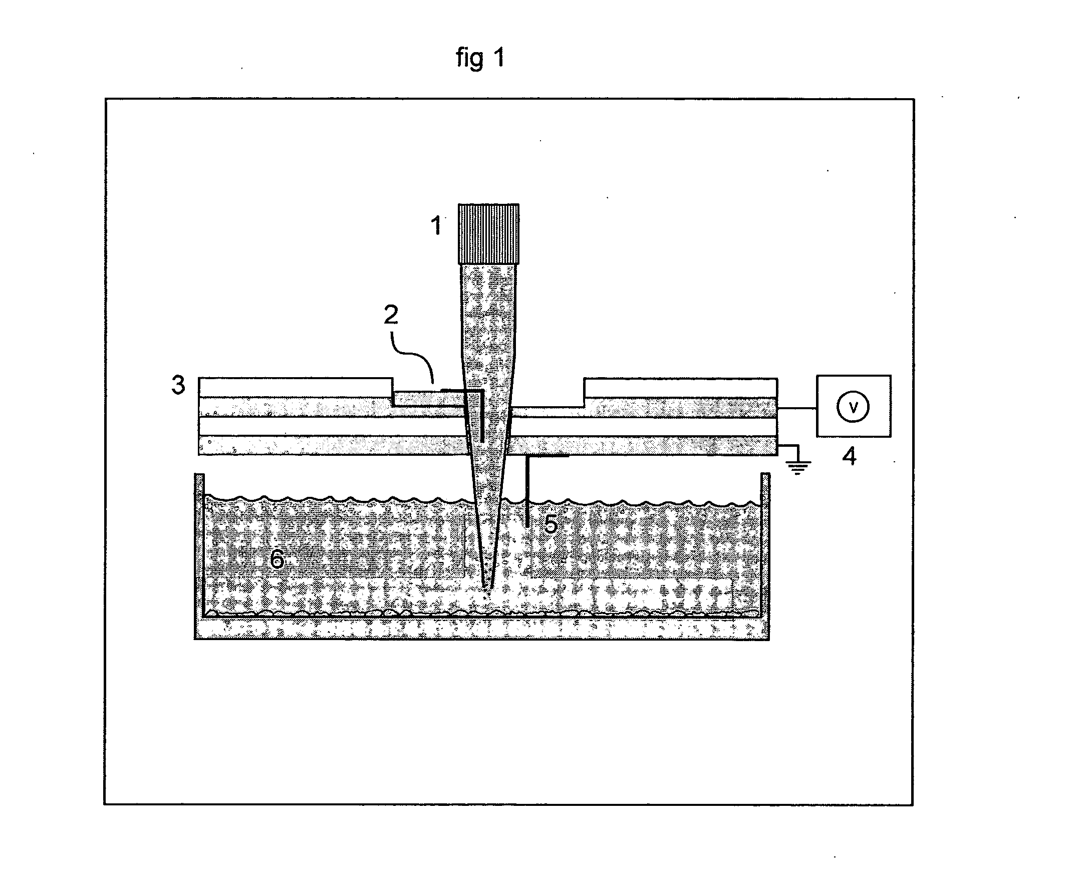
[0311] Electroporation of a Single Spot of Cells in a Confluent Culture galactosidase, PC-12 cells were cultured of standard procedures in conventional petri dishes. The cell culturing media was removed and replaced with a Hepes Saline buffer, pH 7.4, and the cell culture were transferred to the stage of an inverted microscope (Leica, model DMIRB equipped with a x-y translational stage). A tip-electrode manufactured from a conventional pipette tip with a platinum wire inserted horizontally to the interior of the tip, was filled with Hepes-saline buffer supplemented with 500 μM Fluorescein diphosphate (FDP). The tip-electrode was connected to a pulse generator (AM-systems isolated pulse stimulator, model 2100) via an oscilloscope for monitoring of the pulse. A counter electrode in the cell bath was connected to ground and the sign of the potential in the tip-electrode was negative. The outlet of the tip-electrode were positioned at a distance of 50 μm above the cell culture. The cell...

example 2

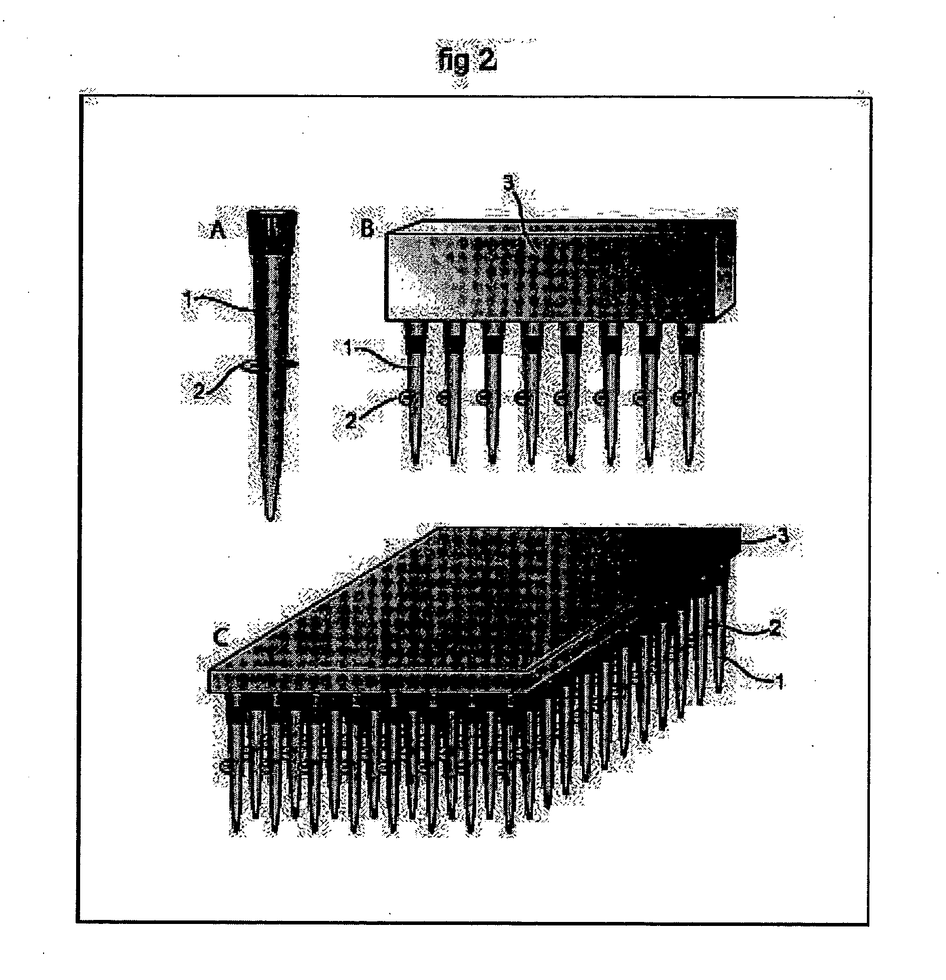
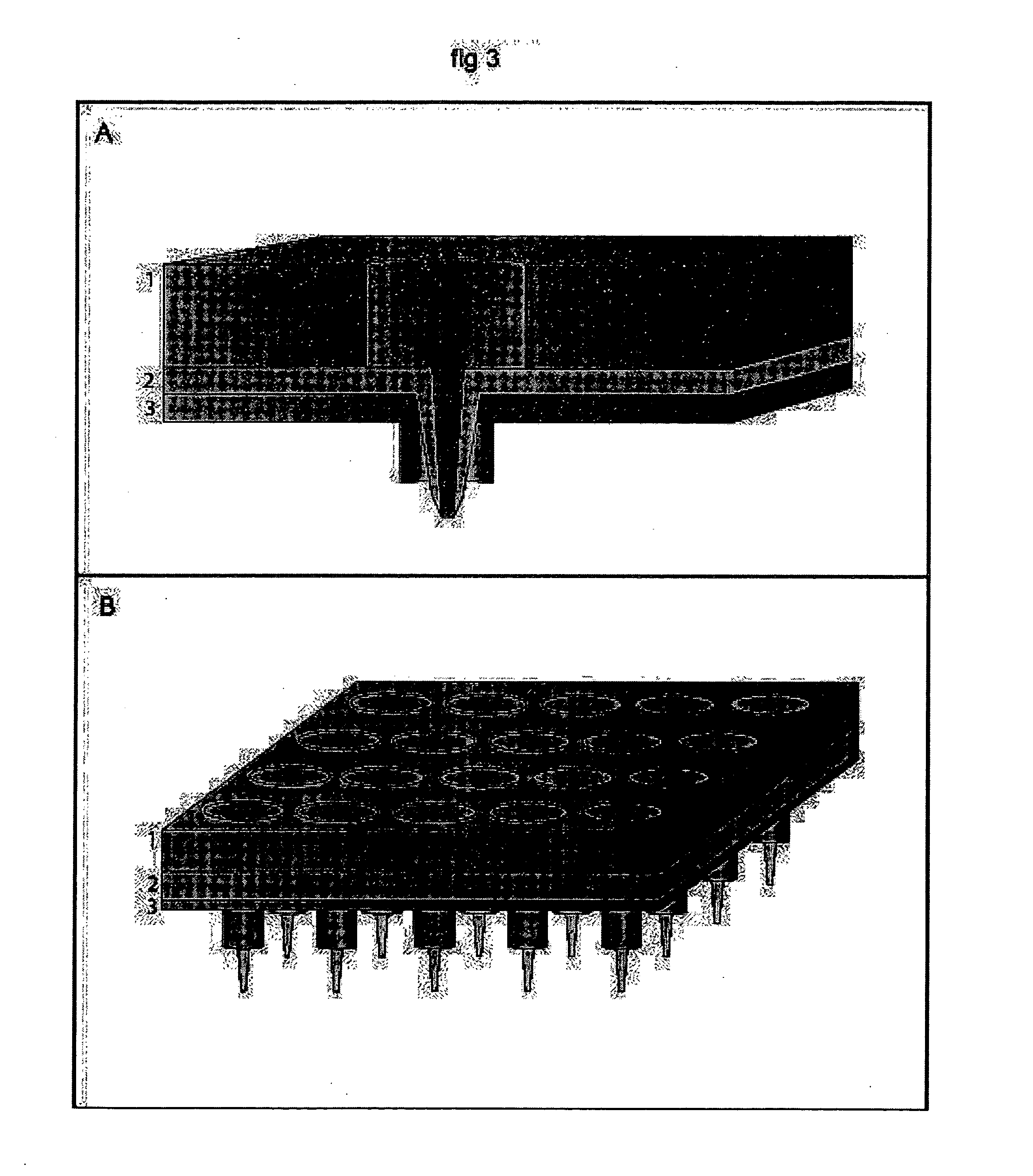
[0312] Parallel Electroporation of Spots in a Confluent Cell Culture PC-12 cells were cultured of standard procedures in conventional petri dishes. The cell culturing media was removed and replaced with a Hepes Saline buffer, pH 7.4, and the cell culture were transferred to the stage of the inverted microscope. Two tip-electrodes, connected to the pulse generator via a single rod inserted through both tips, were filled with Hepes-saline buffer supplemented with 500 μM Fluorescein diphosphate (FDP). One counter electrode was placed in the cell bath and connected to ground. The outlet of the two tip-electrodes were positioned at a distance of approximately 100 μm above the cell culture. Two spots of cells, under the two tip-electrodes, were exposed to 4×10 pulses of 100 ms duration, with a 100 ms delay time between pulses of 4 mA amplitude. FDP was transferred out of the tip-electrodes by electroosmotic forces, no additional pressure or pumping were added to the system. Detection and ...


PUM

PropertyMeasurementUnit
lengthaaaaaaaaaa
lengthaaaaaaaaaa
lengthaaaaaaaaaa

Abstract

The invention provides hollow-tip-electrodes for spatially localized delivery of substances to one or more biological targets present in a population comprising target and non-target molecules, macromolecules, and / or cells. The invention also provides electrode plates for receiving one or more of such tips, tip-electrode plates comprising electrode plates comprising one or more electrode tips, and systems comprising tip-electrodes and containers for containing one or more biological targets, e.g., such as molecules, macromolecules, and / or cells. The invention further provides methods for using such systems and components thereof. In one preferred aspect, the systems are used for spatially confined electroporation of cells and cell structures. The invention facilitates high throughput screening of agents (e.g., such as drugs) that act on intracellular targets.

Description

RELATED APPLICATIONS [0001] This application is a continuation-in-part of U.S. patent application Ser. No. 10 / 325,691, filed Dec. 19, 2002, which is a continuation of U.S. patent application Ser. No. 09 / 557,979, now U.S. Pat. No. 6,521,430, filed Apr. 25, 2000, which is a continuation application of International Application No. PCT / SE98 / 02012, filed Nov. 6, 1998, which claims priority to Swedish Application No. 9704076-0, filed Nov. 6, 1997 and is also a continuation in part of U.S. patent application Ser. No. 10 / 345,107, filed Jan. 15, 2003, which claims priority to U.S. Provisional Application No. 60 / 356,377, filed Feb. 12, 2002. The entireties of these applications are incorporated by reference herein.FIELD OF THE INVENTION [0002] The present invention relates to a method for electroporation and delivery of one or more agents to spatially confined targets, such as populations of cells, single cells, intracellular organelles, and biomolecules. BACKGROUND OF THE INVENTION [0003] D...

Claims


Application Information

Patent Timeline
Patent Type & Authority Applications(United States)
IPC IPC(8): A61N1/30C12N13/00G01N33/543
CPCA61N1/306C12M35/02G01N33/5438G01N33/54366G01N33/54373C12N13/00C12M33/04C12M33/06
Inventor RYTTSEN, FRIDAORWAR, OWELEVIN, MIKAELSAHLIN, ESKILWIGSTROM, JOAKIM
Owner CELLECTRICON
Why Eureka
Try Eureka