Medical instrument for cornea operation

Inactive Publication Date: 2005-03-24
KOKUSAI COMMERCE
View PDF15 Cites 2 Cited by
  • Summary
  • Abstract
  • Description
  • Claims
  • Application Information

AI Technical Summary

Benefits of technology

[0021] Consequently, it is an object of the present invention to provide a medical instrument used in a corneal surgical operation for preparing a complete corneal sheet in a short time and in an easy manner without performing any medication with a medicine which damages the corneal sheet, wherein; the corneal sheet is constructed exclusively of all the layers of the corneal epithelium only; and, the corneal sheet is prepared while enjoying the advantages inherent in the LASEK operation in the corneal refractive correction surgical operation.
[0022] Further, it is another object of the present invention to provide a medical instrument used in a corneal surgical operation for preparing a complete corneal sheet without using any culturing techniques and without suffering from any immune rejection which occurs in a corneal grafting operation conducted between a patient and another person, wherein the corneal sheet is constructed exclusively of all the layers of the corneal epithelium only.
[0024] A medical instrument used in a corneal surgical operation comprising a member positioned in the boundary between a cornea epithelium and a corneal stroma, wherein the member is provided with: a resiliency which makes it possible for a first contact surface of the member to deform along a shape of a contact surface of the corneal stroma in response to a driving force applied to the member in a direction toward a bottom portion of the corneal stroma; and, a strength which makes it possible for a second contact surface of the member to push up lowermost bottom, surface or contact surface of the corneal epithelium wherein the member is driven along the boundary surface between the corneal stroma and the corneal epithelium in response to a thrust applied in a direction along the boundary surface.
[0034] a member positioned in the boundary between a cornea epithelium and a corneal stroma, wherein the member is provided with a resiliency which makes at possible for a first contact surface of the member to deform along a shape of a contact surface of the corneal stroma in response to a driving force applied to the member in a direction toward a bottom portion of the corneal stroma; and, a strength which mares it possible for a second contact surface of the member to push up the lowermost bottom surface or contact surface of the corneal epithelium when the member is driven along the boundary surface between the corneal stroma and the corneal epithelium in response to a thrust applied in a direction along the boundary-surface;
[0041] In a eighth aspect of the present invention, such predetermined inclination angle is 15 degrees. This inclination angle of 15 degrees makes it easy to set the predetermined angle at an angle of 45 degrees in view of an uneven outline or contour of a face of the patient when both the driving force and the thrust are components of a force, wherein: the force is applied to the member at a predetermined angle with respect to a tangential line extending along a direction in which the thrust is applied; and the second contact surface is driven on the contact surface of the corneal stroma.

Problems solved by technology

However, a complete recovery is not expected.
In many cases, such incised wound having been cured or healed results in dim (cloudy) sight.
Such separation is a delicate operation requiring a long time of approximately 10 minutes due to fragility of the corneal epithelium itself.
This replacement or return operation is also delicate operation as well as the above-mentioned separation operation.
On the other hand, the following defects are enumerated: namely, a first defect resides in that a paint or a feeling of physical disorder continues for approximately 4 days or around until the corneal epithelium 51 is regenerated to cover the incised wound; a second defect resides in that the incised wound having been healed often (is apt to) results in dim (cloudy) sight in which a cloud appears under the corneal epithelium; and, a third defect resides in that the operation requires a long period use of a medicine for relief from the cloud the cloudy sight appearing under the corneal epithelium.
On the other hand, defects of this operation are enumerated as follows: namely, a first defect resides in that this operation is more complicated than the PRK operation; a second defect resides in that this operation is more limited in applicability than the PRK operation, wherein an example in poor applicability of this operation is that the microkeratome is suitable for a straight incision use but not suitable for a curved incision use along a curvature of the cornea of the patient, wherein, since a width between an upper and a lower eyelid of an opened eye globe is narrow, it is impossible to set the suction ring; and, a third defect resides in that this operation more suffers from the disease concurrent with the incised wound both during the operation and after the operation than the PRK operation, wherein, once the concurrent disease appears, such concurrent disease tends to become a serious disease.
A fourth defect of this operation resides in, that the corneal stroma 55 is inferior in regeneration ability to the corneal epithelium 51, which results in a very slow healing of the incised wound and also in the non-physiological healing.
However, in the case of the LASEK operation, since the corneal epithelium sheet is subjected to the medication with alcohol, the cells of the epithelium are damaged to such as extent that all the cells are destroyed.
Due to this, the LASEK operation is disadvantageous in that the healing of the incised wound of the corneal epithelium 51 is slow.
As described above, the LASER operation is a delicate operation and requires much more operation time.
Further, in culturing the corneal sheet, a large-scaled facility or installation is required.
This requires much more culturing time.

Method used


Image

  • Medical instrument for cornea operation
  • Medical instrument for cornea operation
  • Medical instrument for cornea operation

Examples


Embodiment Construction

[0067] The best modes for carrying out the present invention will be described using preferred embodiments of the present invention with reference to the accompanying drawings.

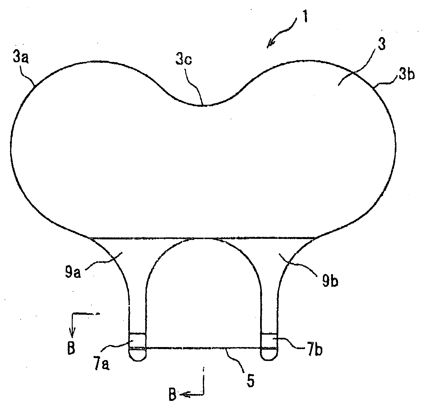
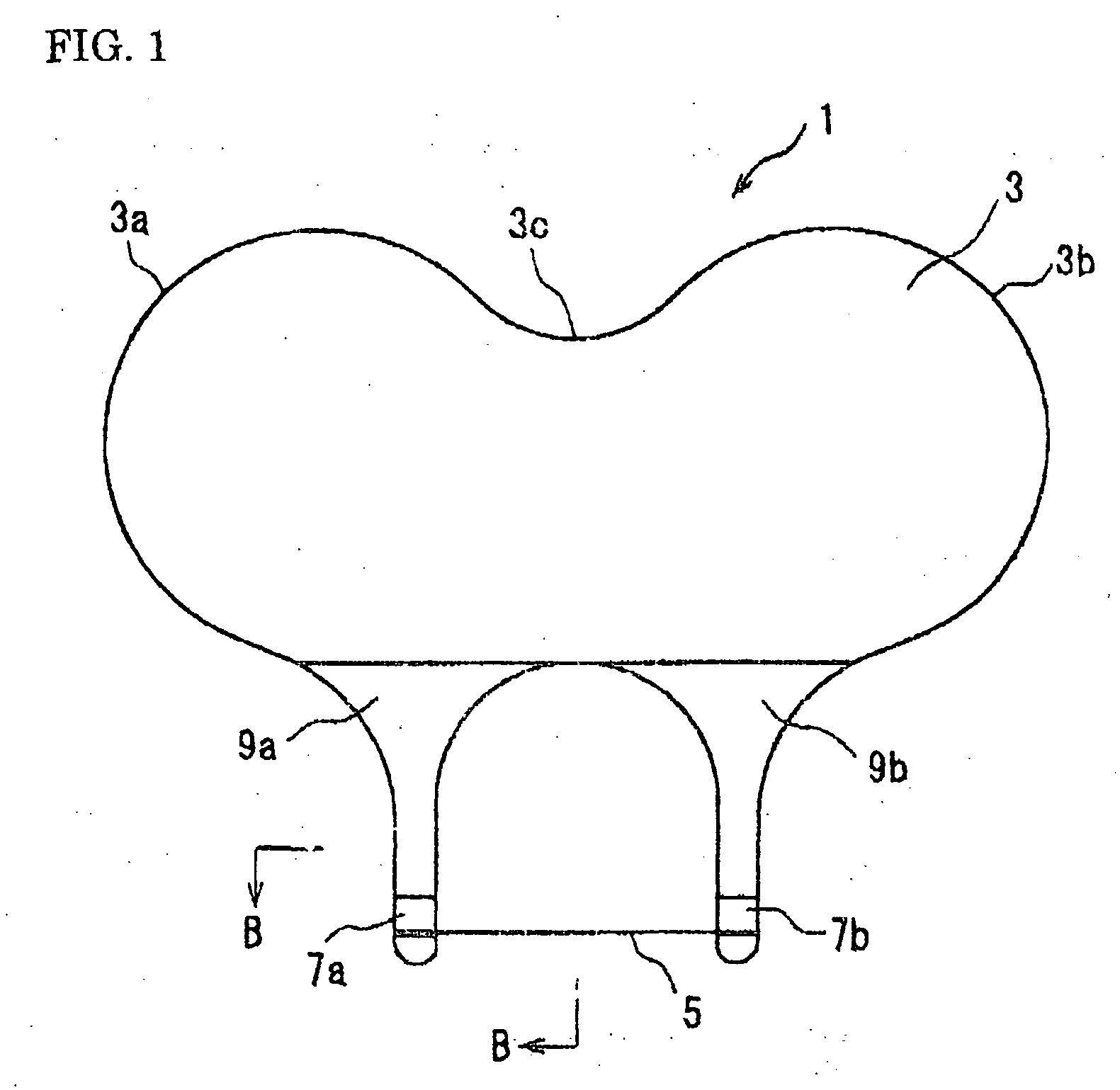
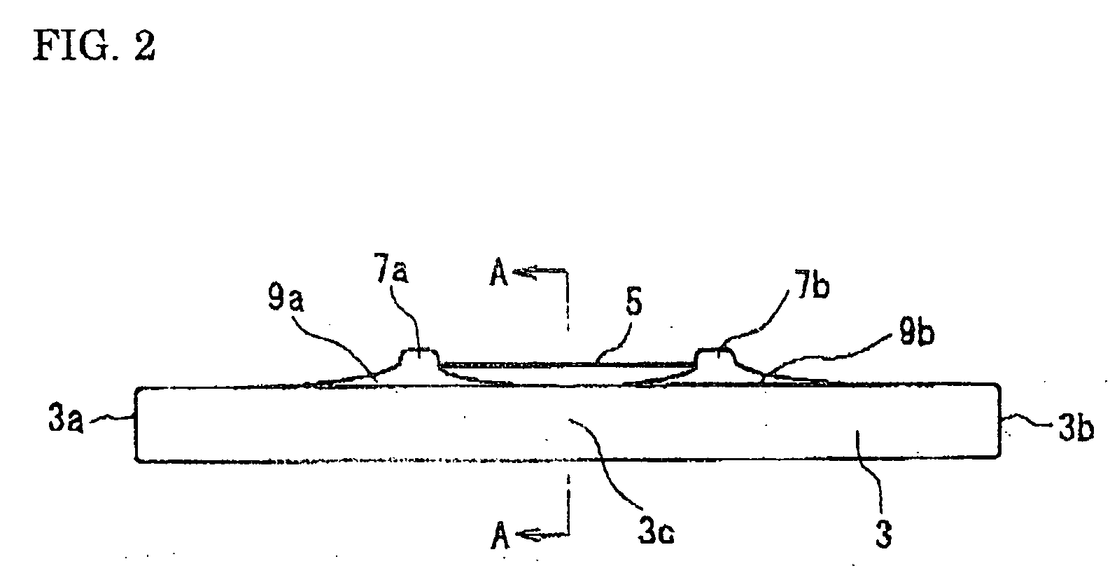
[0068]FIG. 1 is a front view of an instrument for preparing the corneal epithelium sheet, wherein the instrument serves as a medical instrument for carrying out a surgical operation of the cornea according to an embodiment of the present invention. FIG. 2 is a plan view of the instrument for preparing the corneal epithelium sheet shown in FIG. 1. FIG. 3 is a right side view of the instrument for preparing the corneal epithelium sheet shown in FIG. 1. FIG. 4 is a bottom view of the instrument for preparing the corneal epithelium sheet shown in FIG. 1. FIG. 5 is a sectional view, taken along the A-A line of FIG. 2. FIG. 6 is an enlarged view of an essential portion, taken along the line B-B of FIG. 1. FIG. 7 is a sectional view, taken along the line C-C of FIG. 6. FIG. 8 is a sectional view, taken along the lin...


PUM


Abstract

Provided here is a medical instrument, which is employed in a surgical operation of a cornea and capable of preparing a corneal epithelium sheet (including a flap and a cap) in a corneal refractive correction surgical operation or a corneal epithelium grafting operation. The medical instrument employed in the corneal surgical operation of the present invention is provided with a thread-like member 5 which has a diameter D of up to 100 μm and is positioned in the boundary between a corneal epithelium 51 and a corneal stroma 55. The thread-like member 5 is provided with: a resiliency which makes it possible first contact surface 5a of the thread-like member 5 to deform along a shape of a contact surface of a corneal stroma 55 in response to a driving force F applied to the thread-like member 5 in a direction toward a bottom portion of the corneal stroma 55; and, a strength which makes it possible for a second contact surface 5b of the thread-like member 5 to push up the lowermost bottom surface or contact surface of a corneal epithelium 51 when the thread-like member 5 is driven along the boundary surface 61 between the corneal stroma 55 and the corneal epithelium 51 in response to a thrust f2 applied in a direction along the boundary surface 61.

Description

TECHNICAL FIELD [0001] The present invention relates to a medical instrument for carrying out a surgical operation of cornea, and more particularly to a medical instrument used in a corneal refractive correction surgical operation or a corneal epithelium grafting operation. BACKGROUND ART [0002] A cornea is positioned at a starting point of an optical path in visual function to pass and refract an incident light. Consequently, the cornea is an essential organ to realize a sound visual function. [0003]FIG. 19 is a sectional view showing the cornea in construction. [0004] The cornea is constructed of: a corneal epithelium 51 composed of from 6 to 7 layers; Bowman's layer 53; a corneal stroma 55 which forms approximately 80% of the cornea; Descemet's membrane 57 which is the basal membrane of endothelium cells; and, an endothelium composed of hexagonal cells. The cornea has a thickness of approximately 500 μm (L), in which: the corneal epithelium 51 forms an outer layer; and, the endot...

Claims


Application Information

Patent Timeline
IPC IPC(8): A61F9/007A61F9/013
CPCA61F9/007A61F9/0133A61F9/013
Inventor TAKAHASHI, KEIZO
Owner KOKUSAI COMMERCE
Why Eureka
Try Eureka