
Detection of endometrial pathology

a technology of endometrial pathology and detection method, which is applied in the direction of biological material analysis, material analysis, instruments, etc., can solve the problems of low specificity, no routine screening test of practical utility, and carry the risk of uterine perforation or contamination of the cavity, so as to increase the predictive power of the screening assay

Inactive Publication Date: 2005-05-12
STC UNM
View PDF22 Cites 14 Cited by
  • Summary
  • Abstract
  • Description
  • Claims
  • Application Information

AI Technical Summary

Benefits of technology

[0024] In yet another aspect, the invention provides a method for screening a patient or population of patients for endometrial pathology by assaying for the presence of at least one biomarker polypeptide associated with endometrial pathology in a sample obtained from a patient. The biomarker polypeptide is preferably one that is represented by a M / Z peak value in Tables 3, 4, 5, 6 or 7 (Example I). The assay can be a mass spectrometric assay but advantageously can also be an immunoassay, such as a Western blot or an enzyme linked immunosorbent assay (ELISA). A plurality of biomarker polypeptides can be analyzed, thereby increasing the predictive power of the screening assay.

Problems solved by technology

There are currently no routine screening tests of practical utility in detection of endometrial cancer.
Endometrial biopsies and curettings cannot be considered a primary screening tool because they are invasive, can cause cramping and bleeding, and carry risks of uterine perforation or contamination of the cavity by pathogens.
Cytologic evaluation of PAP smears is, however, an insensitive means of endometrial cancer detection and for this reason is not recommended.
Specificity is very low, making this an expensive (during follow up of numerous false positives) as well as insensitive test.
Pre-cancerous and other benign endometrial lesions, such hyperplasia and endometriosis, also pose a significant health risk for women, and convenient screening tests are not available to diagnose these conditions.
Without convenient and easily accessible screening tests for cancer, diagnostic delays will continue to plague the health care system and thwart efforts to detect and treat malignancies in their earliest stages.

Method used


Image

  • Detection of endometrial pathology
  • Detection of endometrial pathology
  • Detection of endometrial pathology

Examples


examples

[0080] The present invention is illustrated by the following examples. It is to be understood that the particular examples, materials, amounts, and procedures are to be interpreted broadly in accordance with the scope and spirit of the invention as set forth herein.

example i

Identification of Peaks Associated With Endometrial Cancer

Summary

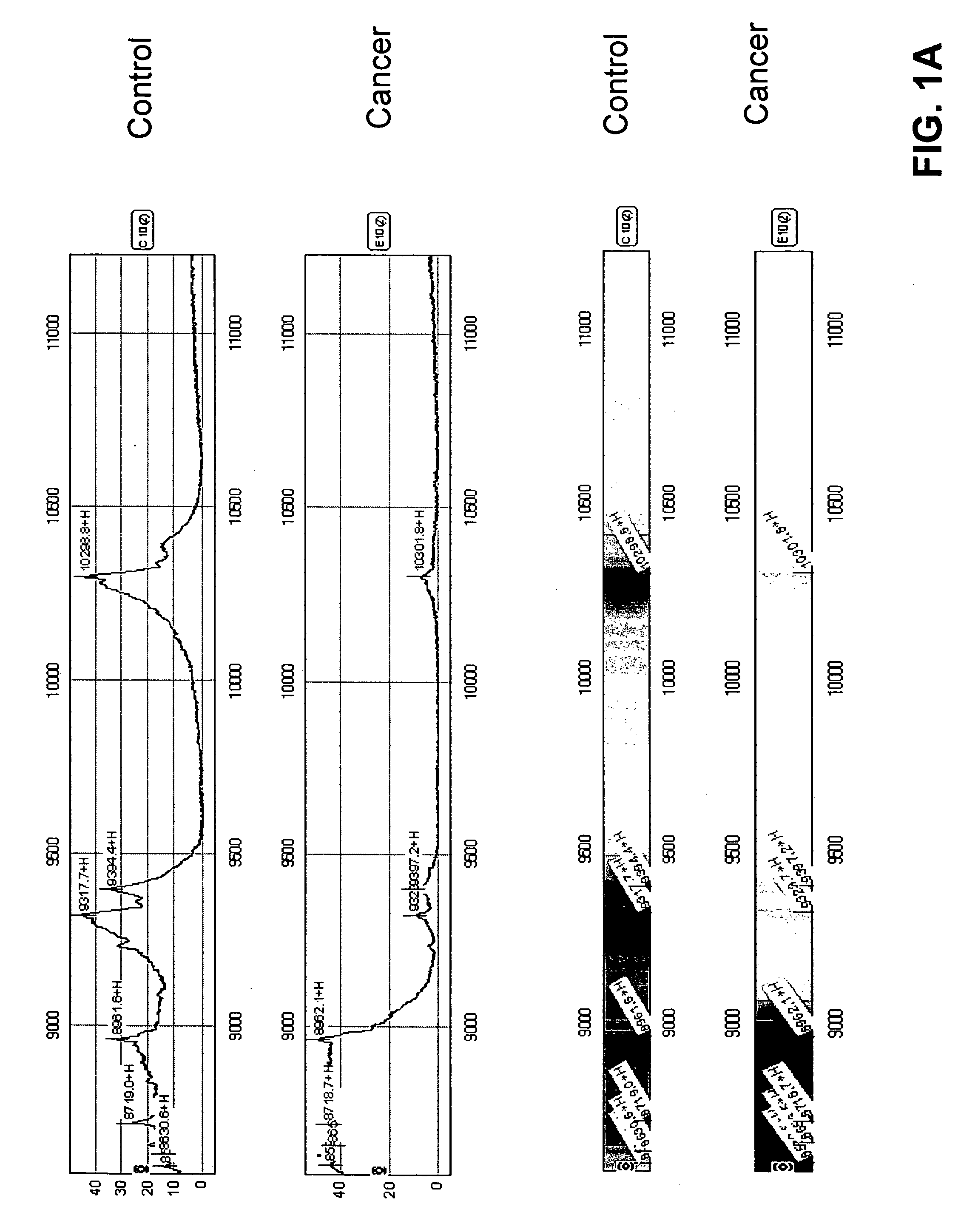
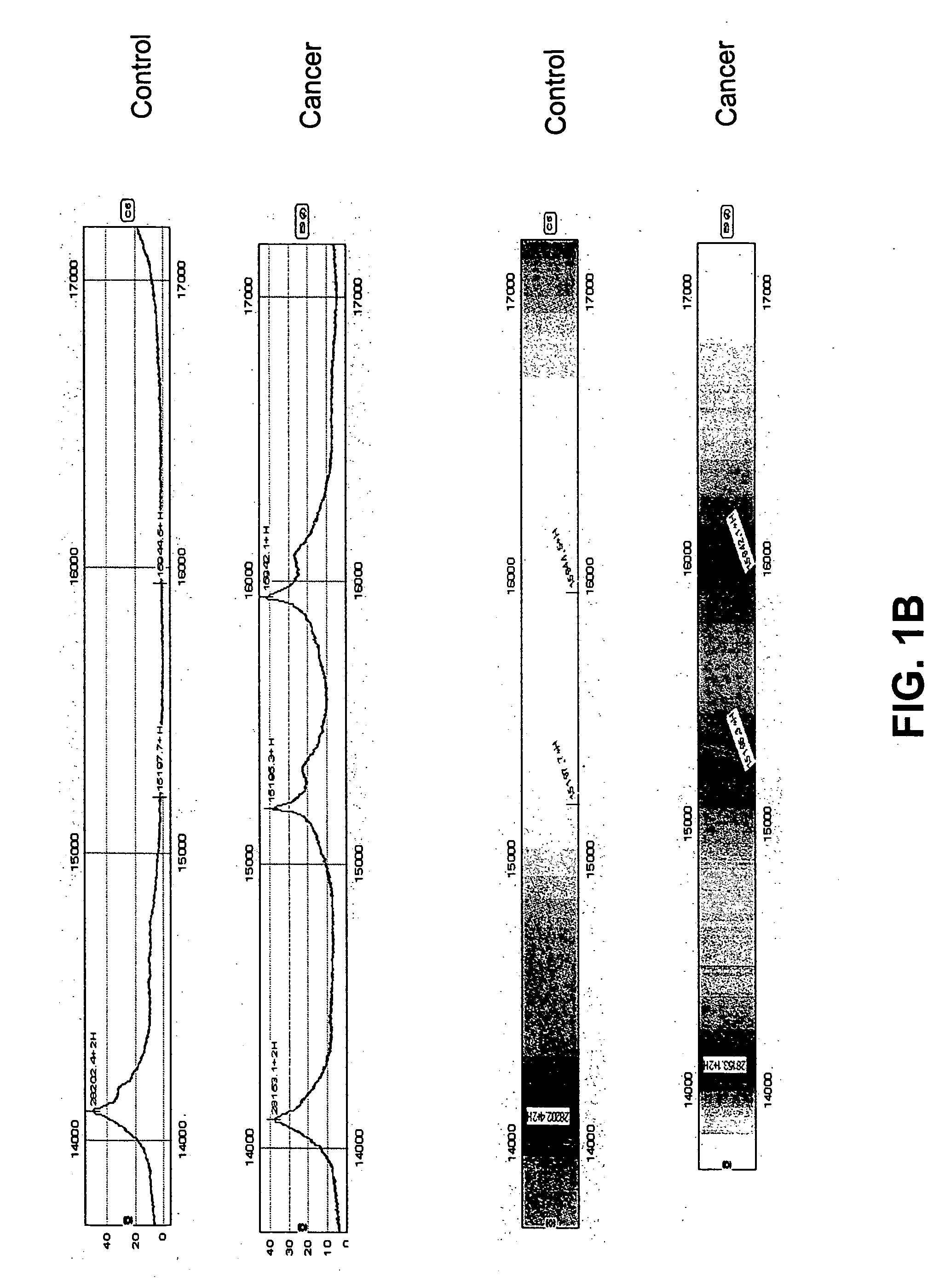
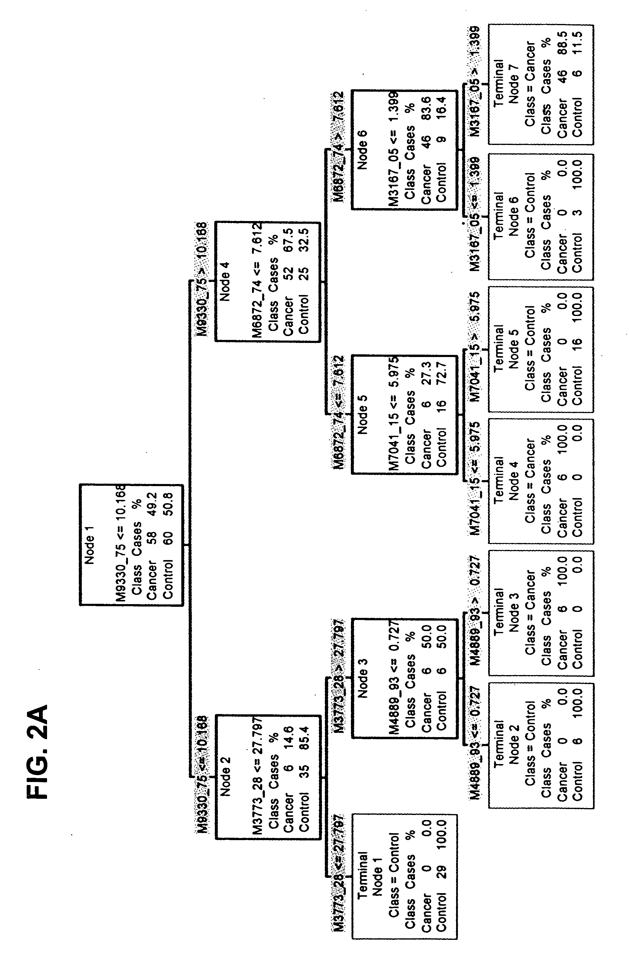
[0081] Mass spectrometry data were collected from serum samples from women with endometrial cancer and from controls. A candidate protein expression profile was identified that appears to distinguish early stage endometrial carcinoma from healthy controls. Peptide spectra were used to create algorithms with high sensitivity and high specificity in discriminating endometrial cancer.

Methods

[0082] Serum was collected in serum separator tubes. It is envisioned that in future work serum will be collected from all patients at diagnosis, at specific times during therapy, 6 months post-therapy, and at recurrence. At collection, the serum is spun in a low speed centrifuge at 1500 rpm for 3 minutes, and the serum is aliquoted into 1 ml cryovials and immediately frozen in liquid nitrogen. Samples will be forwarded to a central tissue collection facility for storage. At the laboratory, samples are thawed, a protease inhibito...

example ii

Identification of Particular Polypeptide Biomarkers

[0119] In Example I, whole serum samples were fractionated using Q Ceramic Hyper DF sorbent columns. Any potential discriminatory biomarkers discovered will have already been assigned to a known pH fraction on the IMAC 30 ProteinChip Array surface. Using this knowledge, a marker can be first purified using Q HyperD F column chromatography, then the pH fraction of interest can be purified to enrich for the biomarker. The fraction containing the marker is purified through a second chromatography step using IMAC HyperCel Spin Columns (Ciphergen, Fremont Calif.), which have a matched chemistry to IMAC 30. The marker is additionally purified, concentrated and desalted using a reversed phase step. Finally the protein of interest is purified using one dimensional SDS PAGE.

[0120] Following in-gel digestion, protein identification can be attempted by MALDI Time of Flight Mass Spectrometry (MALDI-TOF) using an AP Biosystems Voyager Elite in...


PUM


Abstract

Biomarkers and polypeptide profiles for screening, detection, and monitoring of endometrial pathology, including endometrial cancer.

Description

[0001] This application claims the benefit of U.S. Provisional Applications Ser. Nos. 60 / 486,528, filed Jul. 11, 2003, and 60 / 559,932, filed Apr. 6, 2004, each of which is incorporated herein by reference in its entirety.STATEMENT OF GOVERNMENT RIGHTS [0002] This invention was made with government support under grants from the National Institutes of Health, Grant Nos. 1-R24CA883399 and 1-R01CA99908-1. The U.S. government has certain rights in this invention.BACKGROUND OF THE INVENTION [0003] Endometrial cancer is the most frequent invasive gynecologic malignancy and the fourth leading cause of cancer in women. When detected early, outcomes are favorable; nevertheless of the approximately 39,000 new cases of endometrial cancer reported annually in the United States, nearly 7,000 women die of advanced disease. Lifetime endometrial cancer risk in the US is 2.4%. Endometrial cancer has been identified by the National Cancer Institute as an under-studied disease by the recent Progress Re...

Claims


Application Information

Patent Timeline
IPC IPC(8): G01N33/48G01N33/50G01N33/53G01N33/574G01N33/68G06F19/00
CPCG01N33/57442G01N2800/52G01N2800/364G01N33/6851
Inventor LESLIE, KIMBERLY K.MOBARAK, CHARLOTTE D.SMITH, HARRIET O.
Owner STC UNM
Learn More