Magnetic resonance spatial risk map for tissue outcome prediction

a magnetic resonance and spatial risk map technology, applied in the field of brain magnetic resonance imaging, can solve the problem of not taking into account the location-dependent vulnerability to infarction of a voxel, and achieve the effect of improving the tissue outcome prediction of ischemic tissue, enhancing the predictive performance of the model, and increasing the chance of infarction

Inactive Publication Date: 2007-07-19
THE GENERAL HOSPITAL CORP
View PDF4 Cites 40 Cited by
  • Summary
  • Abstract
  • Description
  • Claims
  • Application Information

AI Technical Summary

Benefits of technology

[0019] Another discovery of the present invention is a location parameter that significantly increases the predictive performance of the model. More specifically, a distance parameter is calculated for each voxel that indicates its distance from the acute core lesion. As shown in FIG. 2, for example, an acute DWI region 1 is identified as the core area from which the ischemic injury extends with time. PWI parameters may define a penumbra region 2 which surrounds the core region 1 and which predicts tissue outcome according to prior methods. According to the present invention tissue outcome is also predicted by its distance from the core region 1. The voxel 3 for example, is located a distanced d1 from the core 1, whereas the voxel 4 is located a distance d2 from the core lesion 1. Because voxel 4 is much closer to the core lesion 1 than voxel 3, the location parameter d will predict a higher chance of infarction for point 4 than point 3.
[0020] A general object of the invention is to provide an MR imaging method that improves the tissue outcome prediction of ischemic tissue. It has been discovered that the acute region identified by DWI imaging can, and usually does, differ from the acute region identified by PWI imaging. One of the two regions will be smaller than the other and this smaller region defines the core region where both measurement techniques agree that infarction will occur. At voxels located outside this core region outcome is less certain and the present invention adds location parameters to the model to improve the outcome prediction.

Problems solved by technology

A limitation of prior models is that they fail to take into consideration the location-dependent vulnerability to infarction of a voxel.

Method used


Image

  • Magnetic resonance spatial risk map for tissue outcome prediction
  • Magnetic resonance spatial risk map for tissue outcome prediction
  • Magnetic resonance spatial risk map for tissue outcome prediction

Examples


Embodiment Construction

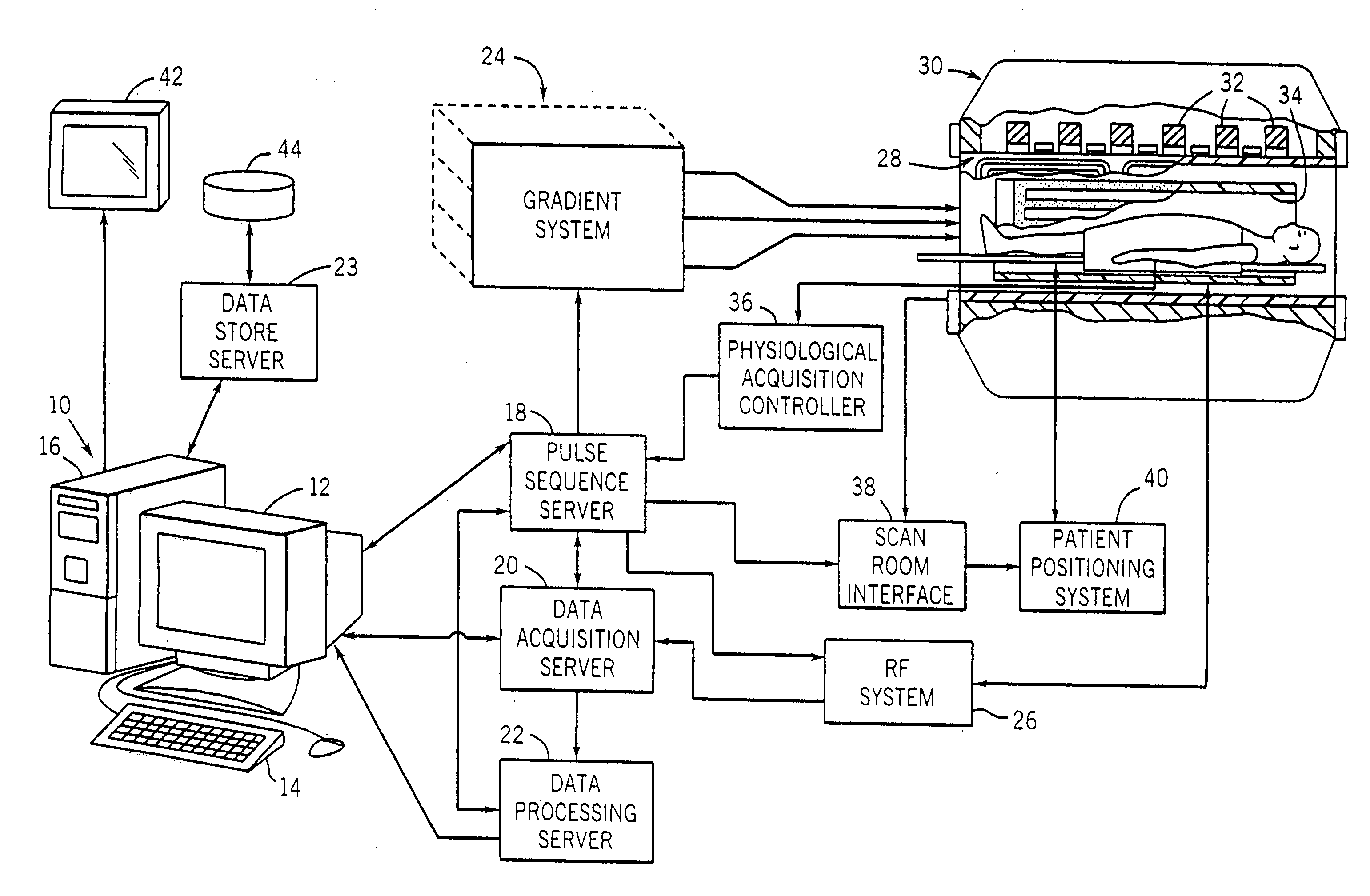
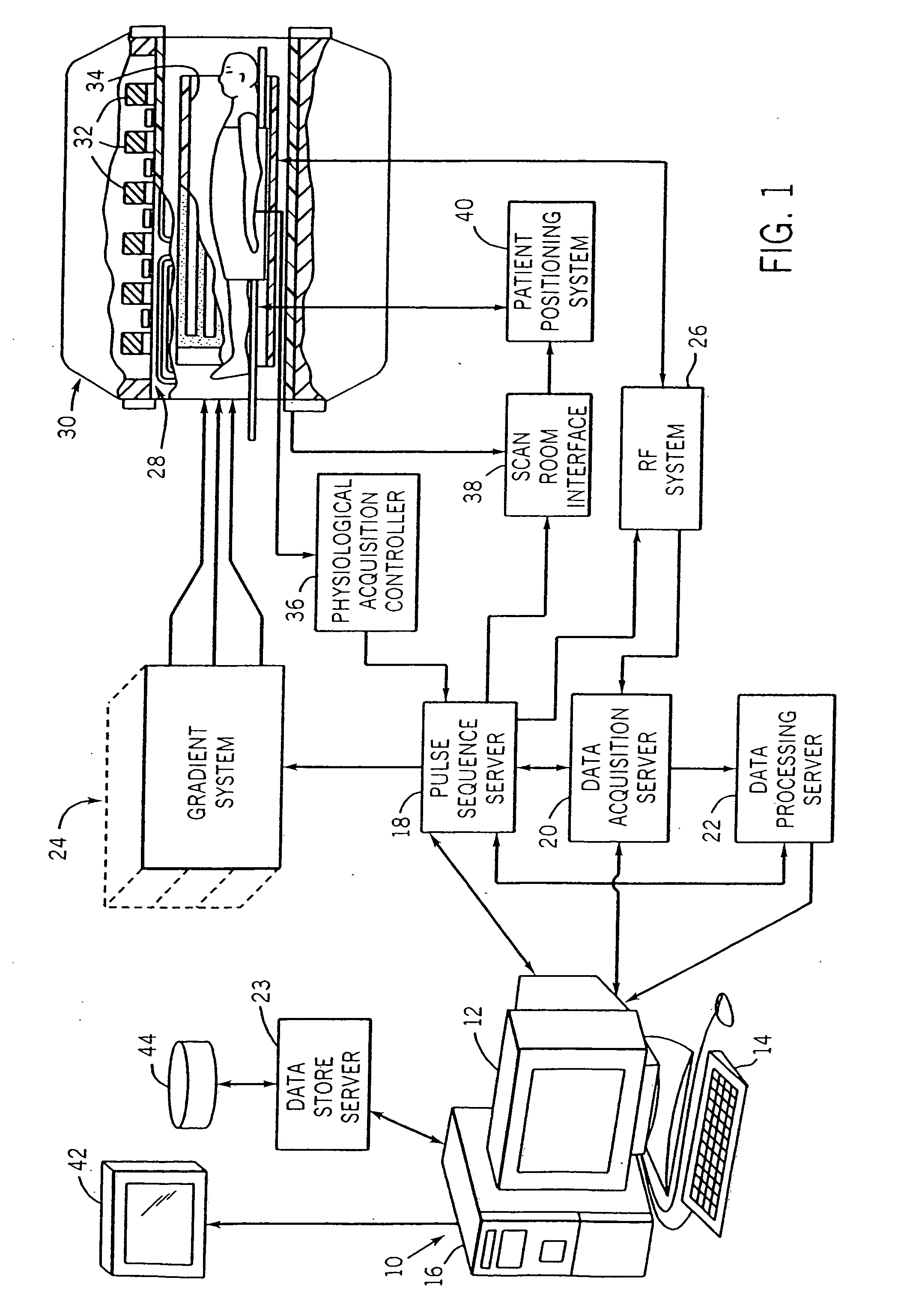
[0026] Referring particularly to FIG. 1, the preferred embodiment of the invention is employed in an MRI system. The MRI system includes a workstation 10 having a display 12 and a keyboard 14. The workstation 10 includes a processor 16 which is a commercially available programmable machine running a commercially available operating system. The workstation 10 provides the operator interface which enables scan prescriptions to be entered into the MRI system.

[0027] The workstation 10 is coupled to four servers: a pulse sequence server 18; a data acquisition server 20; a data processing server 22, and a data store server 23. In the preferred embodiment the data store server 23 is performed by the workstation processor 16 and associated disc drive interface circuitry. The remaining three servers 18, 20 and 22 are performed by separate processors mounted in a single enclosure and interconnected using a 64-bit backplane bus. The pulse sequence server 18 employs a commercially available mi...


PUM


Abstract

Diffusion weighted images and perfusion weighted images are acquired with an MRI system and used to produce low b, DWI, ADC, CBV, CBF, and MTT images of brain tissues following an ischemic event. These MRI physiological measurements are input along with a spatial location measurement to a generalized linear model that predicts the outcome of tissues surrounding a lesion.

Description

CROSS-REFERENCE TO RELATED APPLICATIONS [0001] This application is based on U.S. Provisional patent application Ser. No. 60 / 678,434 filed on May 6, 2005 and entitled “Method For Predicting Tissue Outcome in Acute Human Stroke.”STATEMENT REGARDING FEDERALLY SPONSORED RESEARCH [0002] This invention was made with government support under Grant No. 5R01NS038477-07 awarded by the National Institute of Health. The United States Government has certain rights in this invention.BACKGROUND OF THE INVENTION [0003] The field of the invention is nuclear magnetic resonance imaging (MRI) methods and systems. More particularly, the invention relates to MR imaging of the brain. [0004] When a substance such as human tissue is subjected to a uniform magnetic field (polarizing field B0), the individual magnetic moments of the spins in the tissue attempt to align with this polarizing field, but precess about it in random order at their characteristic Larmor frequency. If the substance, or tissue, is sub...

Claims


Application Information

Patent Timeline
Patent Type & Authority Applications(United States)
IPC IPC(8): A61B5/05
CPCG01R33/56341G06T2207/30016G06T7/0012
Inventor MENEZES, NINASORENSEN, ALMA GREGORY
Owner THE GENERAL HOSPITAL CORP
Why Eureka
Try Eureka