Catheter device

a catheter and tube technology, applied in the field of catheters, can solve the problems of atrial flutter, negative affecting the stimulus conduction capability of the ventricle myocardium, etc., and achieve the effect of simple fashion and safe and purposeful working

Inactive Publication Date: 2005-07-07
SIEMENS HEALTHCARE GMBH
View PDF7 Cites 146 Cited by
  • Summary
  • Abstract
  • Description
  • Claims
  • Application Information

AI Technical Summary

Benefits of technology

[0008] The invention is based on an object of specifying a catheter device which enables the doctor to obtain information from the treatment area in simple fashion, permitting safe and purposeful working.
[0010] The inventive proposed integration of an imaging device in the region of the tip of the catheter allows the doctor particularly advantageously to display the area under examination at high resolution, for example on a monitor, i.e. for example areas of scarred tissue or the delimitation thereof from healthy tissue, i.e. consequently to obtain an internal view of the heart, atrium, etc. Simultaneously he can undertake the ablation using the same catheter, i.e. necrotize the area of scared tissue displayed to him by imaging, using the high-frequency application.
[0016] As described, rotating light decoupling is generally used in the OCT device, a single light-conducting fiber being used. The light decoupling takes place laterally on the catheter, i.e. essentially perpendicular to the catheter's longitudinal axis. However, it is also conceivable to route a bundle of several individual fibers to the tip of the catheter and to place a collimator upstream of the individual fibers to ensure that the light emitted is reflected precisely to that fiber from which the light was emitted. This embodiment enables the OCT light to be coupled out and coupled in at the tip, i.e. in an extension of the catheter's longitudinal axis.

Problems solved by technology

A heart attack often leaves scars, in particular in the ventricle, which negatively affect the stimulus conduction capability in the myocardium of the ventricle.
Particularly in this region containing only partially functionally impaired cells there is a risk that stimulation current conduction is insufficient, resulting in a continuous pulse being emitted which can lead to an atrial flutter.

Method used


Image

  • Catheter device
  • Catheter device

Examples


first embodiment

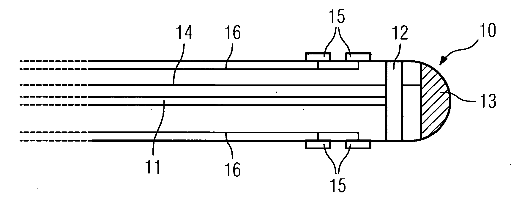
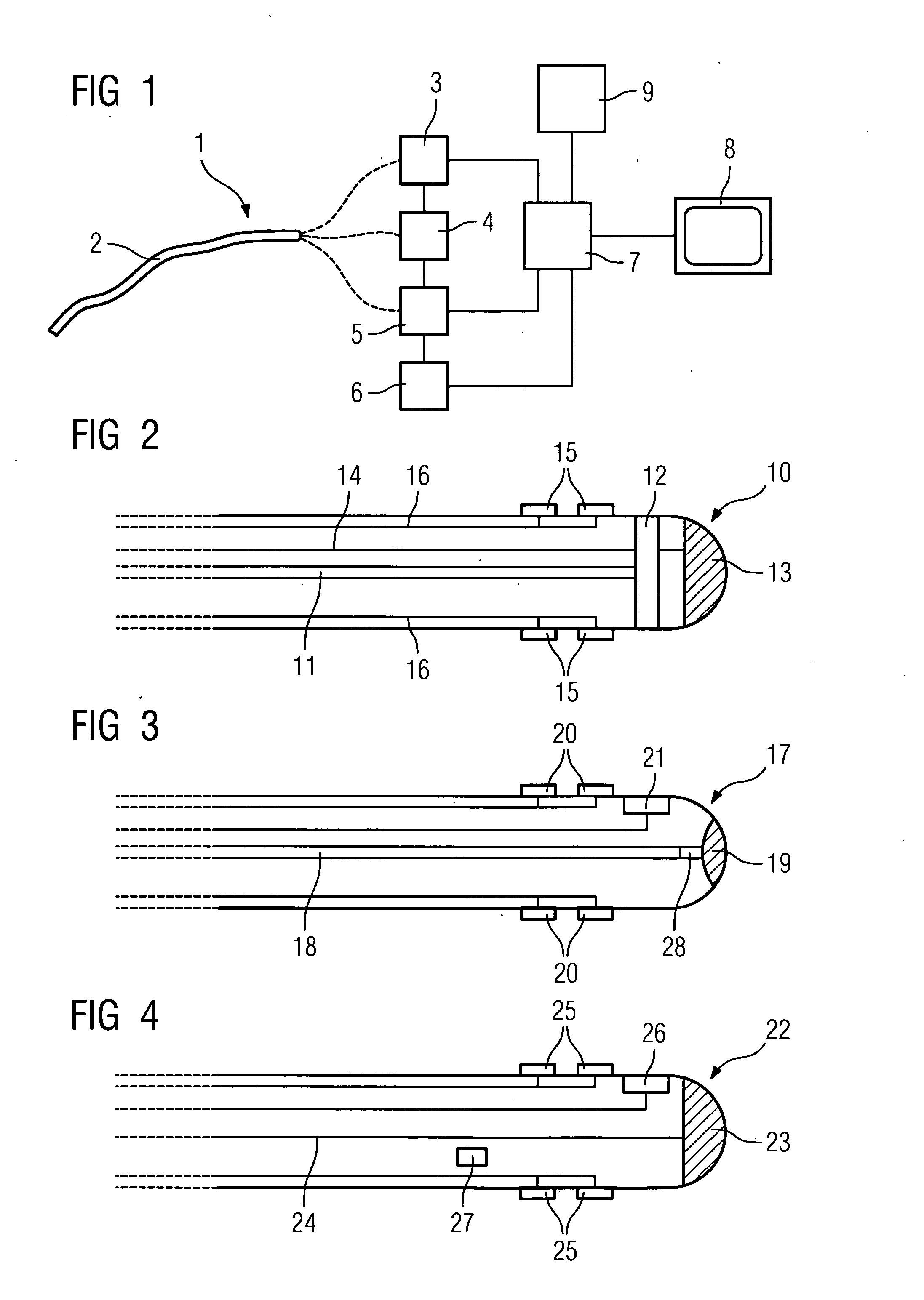
[0032] In the exemplary embodiment, FIG. 2 shows the tip 10 of a catheter of a first embodiment in detail. A thin optical fiber guide 11 is shown, via which the light on which OCT imaging is based is supplied, or the recorded reflected light is coupled out. The light is supplied and the decoupled light is processed using the device 3. A window 12 in the region of the tip of the catheter is provided for this purpose, from which the light is coupled out vertical to the longitudinal axis of the catheter. To this end the optical fiber guide 11 is designed accordingly, as is known. The decoupled light beam rotates, so that an annular image of the area adjacent to the tip is captured.

[0033] An ablation electrode 13 is further provided, which is fed via a power feed line 14. This enables adjacent tissue to be necrotized using high-frequency pulses of current.

[0034] Furthermore a plurality of electrodes 15 is provided, which are arranged on the outside of the catheter and can be controlled...

second embodiment

[0036]FIG. 3 shows another tip 17 of the catheter of a Here too an optical fiber 18 is provided, but it is a fiber bundle of many individual light guides rather than a single fiber. The light is coupled in and out via an OCT lens 19 arranged in the tip in the longitudinal direction of the catheter. Upstream of this is a collimator 20, which ensures that the light emitted by a fiber was reflected precisely to the one from which the light was emitted. This ensures that the OCT imaging is not impaired with this type of “longitudinal coupling in and out”.

[0037] Otherwise electrodes 20 and the ablation device 21 are also provided, the latter being positioned laterally here. Different local positioning options are of course also conceivable here.

[0038] Finally FIG. 4 shows another tip 22 of the catheter. In this case, instead of an OCT imaging device an ultrasound device comprising an ultrasonic head 23 arranged in the tip with a corresponding signal line 24 is provided, via which ultra...


PUM


Abstract

Catheter device comprising a catheter (2), particularly an intravascular catheter, for insertion into an organ or vessel of the human or animal body, with a device for ablation of the adjacent organ tissue or vessel tissue using high-frequency currents in the region of the tip of the catheter, a device (3, 11, 12, 18, 19, 23, 24) being integrated for capturing images of the organ or vessel in the region of the tip of the catheter (10, 17, 22).

Description

CROSS REFERENCE TO RELATED APPLICATIONS [0001] This application claims priority to the German application No. 10355275.8, filed Nov. 26, 2003 which is incorporated by reference herein in its entirety. FIELD OF INVENTION [0002] The invention relates to a catheter device comprising a catheter, in particular an intravascular catheter, for insertion into an organ or vessel of the human or animal body, with an ablation device comprising an ablation electrode arranged in the region of the tip of the catheter for ablation of the adjacent organ tissue or vessel tissue using high-frequency currents, an imaging device to capture images of the organ or vessel in the region of the tip of the catheter, as well as an ECG device to record an ECG comprising one or more electrodes provided in the area of the tip of the catheter to derive an ECG, as known for example from U.S. Pat. No. 6,047,218. BACKGROUND OF INVENTION [0003] Ablation catheters, i.e. catheters with a device for tissue ablation by ne...

Claims


Application Information

Patent Timeline
Patent Type & Authority Applications(United States)
IPC IPC(8): A61B1/00A61B5/00A61B5/055A61B5/296A61B8/12A61B18/12A61B18/14A61B19/00
CPCA61B5/0066A61B5/042A61B5/6852A61B2018/00839A61B18/1492A61B19/5212A61B8/12A61B90/361A61B5/283
Inventor KLEEN, MARTINRAHN, NORBERT
Owner SIEMENS HEALTHCARE GMBH
Why Eureka
Try Eureka