Mesenchymal precursor cell and use thereof in the repair of bone defects and fractures in mammals
a precursor cell and mesenchymal technology, applied in the field of mammals mesenchymal precursor cells and their use in the repair of bone defects and fractures, to achieve the effect of increasing the rate of haemopoiesis
- Summary
- Abstract
- Description
- Claims
- Application Information
AI Technical Summary
Benefits of technology
Problems solved by technology
Method used
Image
Examples
examples 1-3
Results
Example 1
Isolation and Purification of STRO-1+ BM MPC
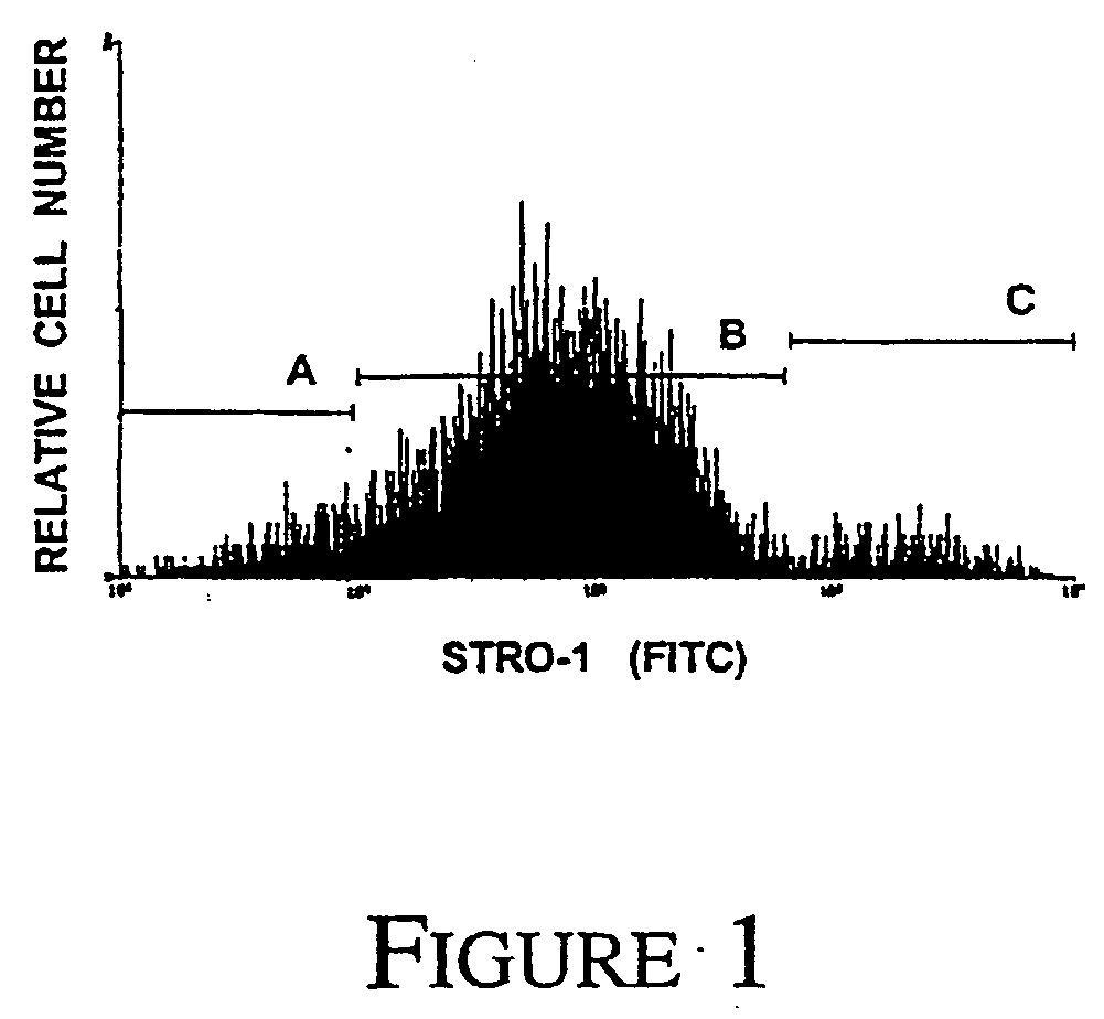
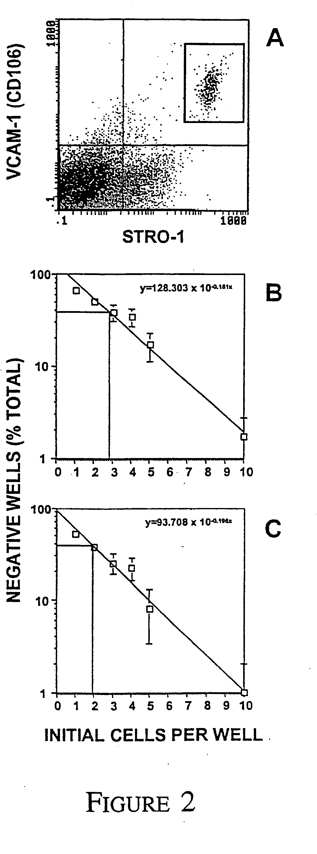
[0096] We have previously demonstrated the effectiveness of MACS to isolate and enrich for MPC from aspirates of human BM based on the cell surface expression of the STRO-1 antibody [Gronthos and Simmons, 1995; Gronthos et al., 1998]. In the present study, flow cytometric analysis of MACS isolated STRO-1+ BMMNC cells demonstrated a heterogeneous pattern of expression spanning over four logs in fluorescence intensity (FIG. 1). Single-color FACS was subsequently employed to sort the STRO-1+ BMMNC fraction into three subsets; STRO-1 dull STRO-1intermediate and STRO-1bright Clonogenic assay for CFU-F in the different sorted STRO-1+ subpopulations demonstrated that the majority of the MPC were contained within the STRO-1bright cell fraction. There was a 900 fold increase in the incidence of CFU-F in the STRO-1bright population when compared to unfractionated BMMNC (Table 1) demonstrating that BM MPC contained a high copy numb...
example 2
Characterization of Purified BM MPC
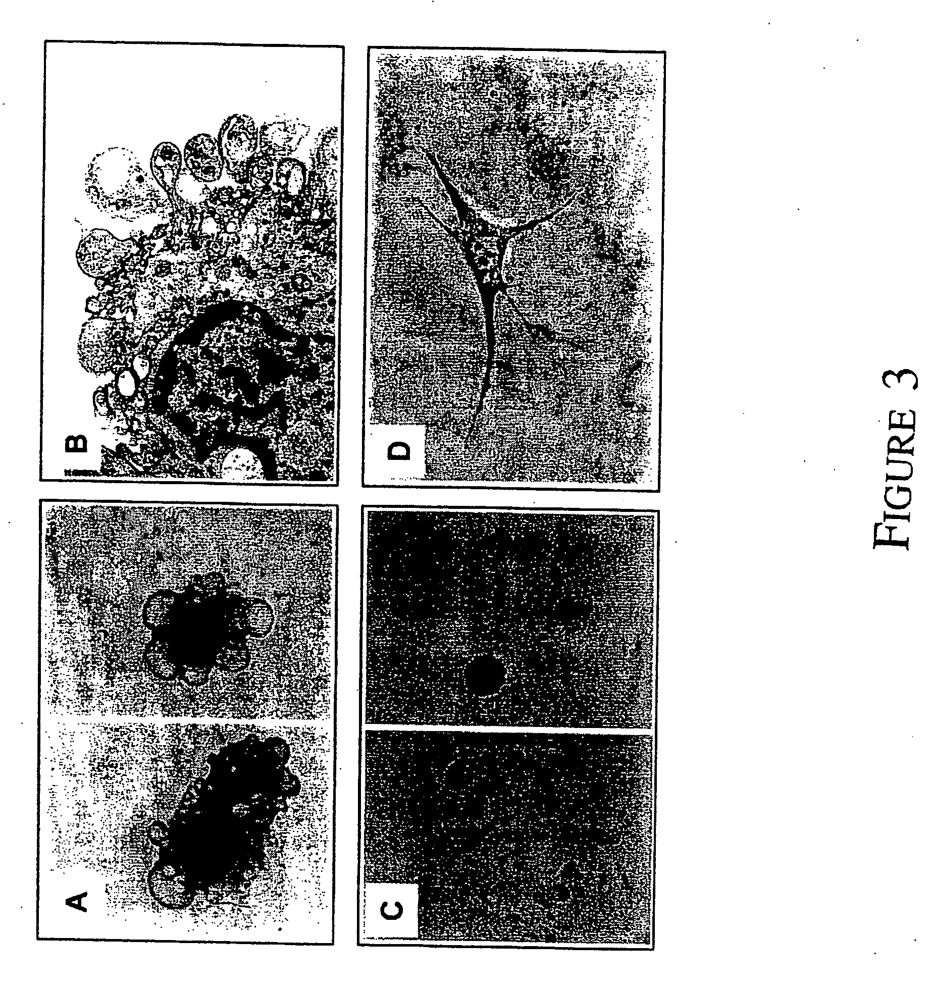
[0098] Morphological examination of freshly sorted STRO-1bright / VCAM-1+ cells was carried out by transmission electron microscopy. Purified BM CFU-F appeared to be a homogeneous population of large cells containing many cytoplasmic processes and a large nucleous with an open chromatin structure (FIG. 3). To determine the cell cycling status of the CFU-F population in aspirates of BM the MACS isolated STRO-1+ BMMNC fraction was further incubated with the cell cycling specific antigen Ki-67 [Gerdes et al., 1984; Wersto et al., 1988]. Two color flow cytometric analysis revealed that the STRO-1bright subset which contained the CFU-F population lacked co-expression of the Ki-67 antigen demonstrating that these cells are non-dividing in vivo (FIG. 4A). Telomerase activity was examined in cell extracts from sorted and cultured candidate stromal progenitor cell populations by a modified TRAP assay. Telomerase activity was present in all fractions includin...
example 3
The Developmental Potential of BM MPC CLONES In Vitro and In Vivo
[0102] Bone marrow CFU-F clones were established from STRO-1bright / VCAM-1+ sorted cells from three individual BM donors. At day 4 of culture, single clonogenic clusters were identified and expanded by subculture. Half of the cells from the first passage were taken from each clone and cultured under osteogenic growth conditions as described above. The osteogenic potential of ninety CFU-F clones was assessed where a von Kossa positive mineralised matrix formed in all of the ninety clones. However, only a proportion (38%±15SEM, n=3) of the same clones gave rise to clusters of lipid containing oil red-O positive adipocytes demonstrating the bi-potential of the CFU-F population in vitro.
[0103] Half the cells from a representative 46 clones were subcultured and expanded for several weeks, then seeded into porous HA ceramic cubes and implanted subcutaneously into SCID mice for a period of 8 weeks as previously described [Ha...
PUM
| Property | Measurement | Unit |
|---|---|---|
| molecular weight | aaaaa | aaaaa |
| molecular weight | aaaaa | aaaaa |
| pH | aaaaa | aaaaa |
Abstract
Description
Claims
Application Information
 - R&D
- Intellectual Property
- Life Sciences
- Materials
- Tech Scout
- Unparalleled Data Quality
- Higher Quality Content
- 60% Fewer Hallucinations
Browse by: Latest US Patents, China's latest patents, Technical Efficacy Thesaurus, Application Domain, Technology Topic, Popular Technical Reports.
© 2025 PatSnap. All rights reserved.Legal|Privacy policy|Modern Slavery Act Transparency Statement|Sitemap|About US| Contact US: help@patsnap.com


