
Electronic scan type ultrasound diagnostic instrument

a diagnostic instrument and ultrasound technology, applied in the field of ultrasound diagnostic instruments, can solve the problems of reducing maneuverability, affecting the operation of the patient, and affecting the patient's comfort, so as to reduce the diameter of the signal line cabl

Inactive Publication Date: 2005-07-28
FUJI PHOTO OPTICAL CO LTD
View PDF8 Cites 18 Cited by
  • Summary
  • Abstract
  • Description
  • Claims
  • Application Information

AI Technical Summary

Benefits of technology

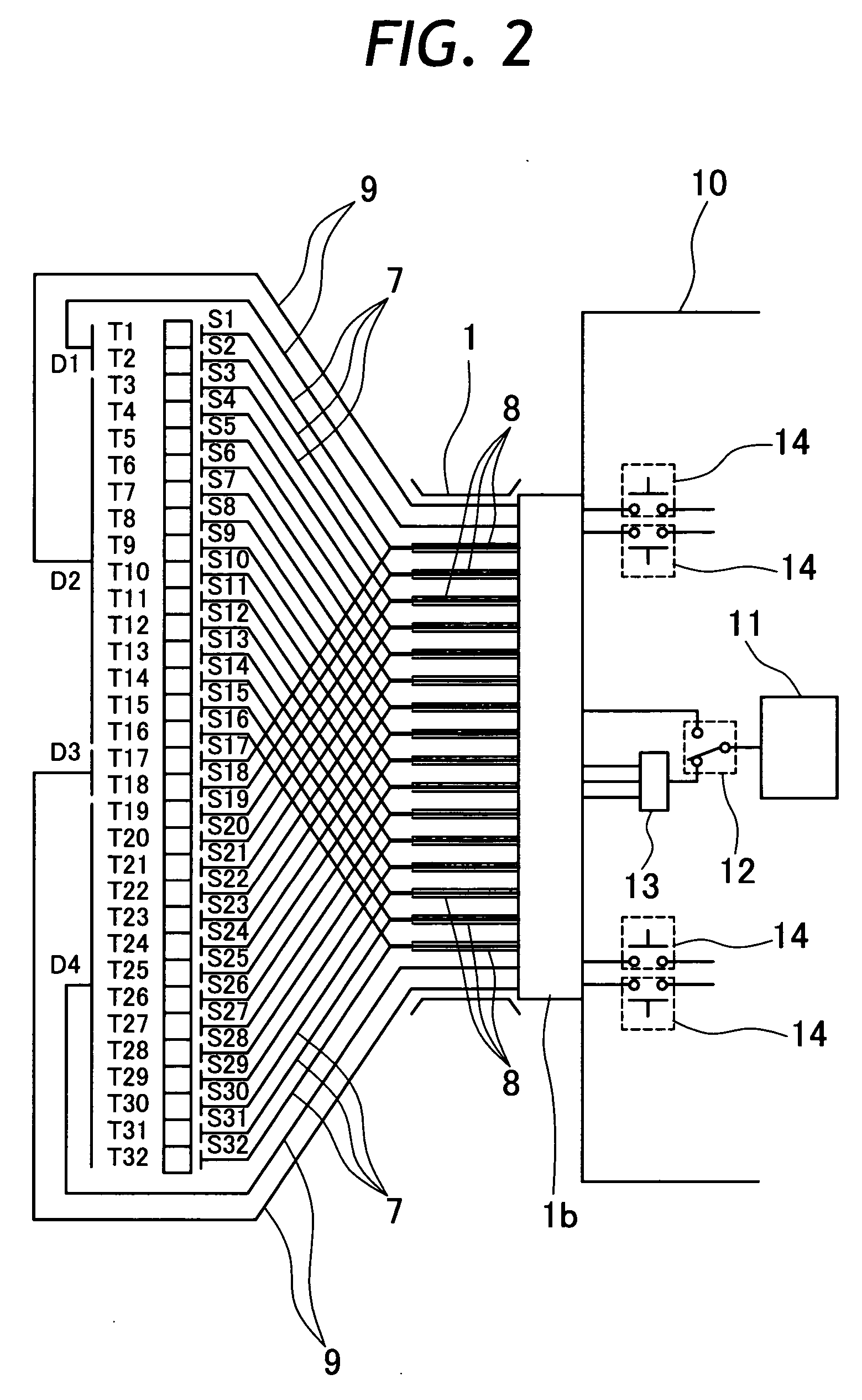
[0009] In view of the foregoing situations, it is an object of the present invention to provide an electronic scan type ultrasound diagnostic instrument with a wiring system using a drastically reduced number of wires to and from transducer elements of an ultrasound transducer, permitting to reduce the diameter of a cable of signal lines.

Problems solved by technology

In this connection, in the case of an ultrasound transducer which is constituted by a large number of transducer elements as mentioned above, there has been a problem, in addition to complication in construction, in that it is inevitable to provide a cable of a large diameter containing a bundle of a large number of hard-wires for making wring connections to and from the respective transducer elements.
Let alone transmission of ultrasound pules from outside a patient's body, the use of a thick cable is undesirable for an ultrasound probe having an ultrasound transducer at the fore end of an insert portion to be introduced into a body cavity of a patient, because the thick cable will not only degrade maneuverability of the probe at the time of insertion into a patient's body but cause greater pains on the part of the patient.
The above-mentioned prior art wiring system succeeded in reducing the number of wires, but still has a problem that the use of switching elements for the respective signal lines results in an ultrasound transducer which is more complicate in construction and larger in size.
Therefore, if the ultrasound transducer is incorporated into an insertion type ultrasound diagnostic instrument, a fore end portion of an insertion tube will become too bulky in consideration of maneuverability of the instrument at the time of insertion into a body cavity.

Method used


Image

  • Electronic scan type ultrasound diagnostic instrument
  • Electronic scan type ultrasound diagnostic instrument
  • Electronic scan type ultrasound diagnostic instrument

Examples


Embodiment Construction

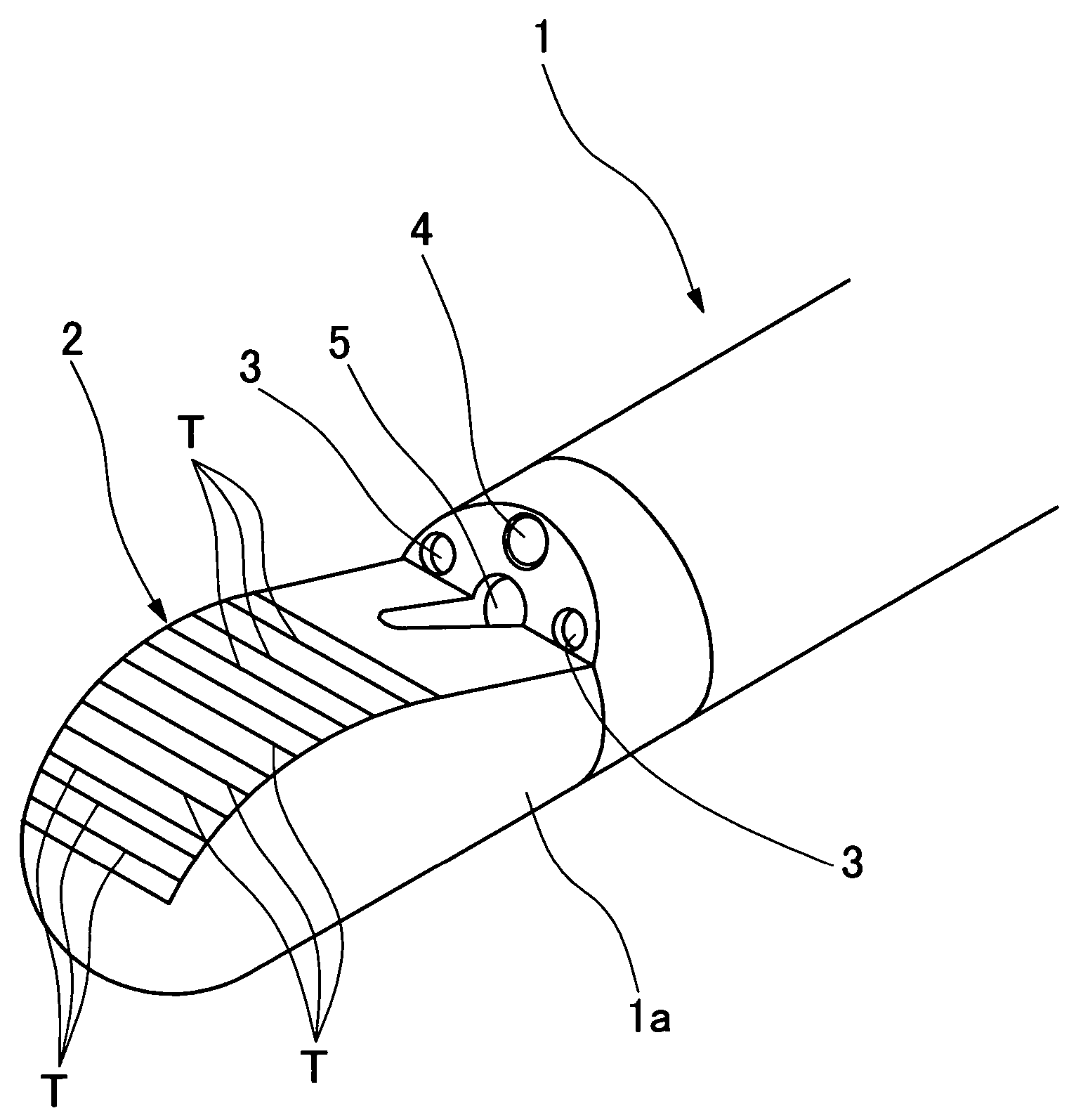
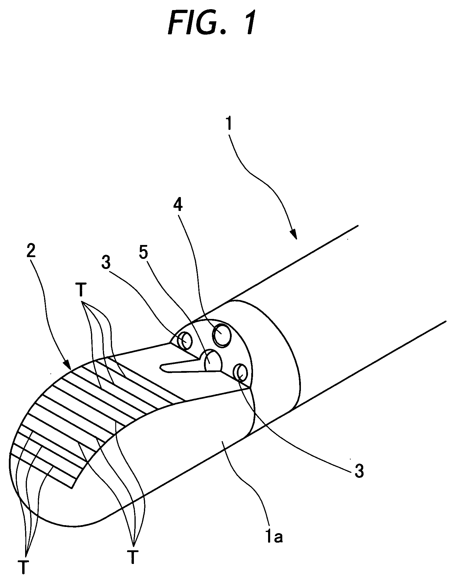
[0031] Hereafter, the present invention is described more particularly by way of its preferred embodiments, with reference to the accompanying drawings. Referring first to FIG. 1, there is shown a fore end portion of an insertion tube of an ultrasound endoscope embodying the ultrasound diagnostic instrument according to the present invention. Application of the ultrasound diagnostic instrument according to the present invention is not limited to ultrasound probes or endoscopes. Namely, the ultrasound diagnostic instrument of the invention can be applied not only as an insertion type diagnostic instrument which makes intracavitary scans like an endoscope, but also as an external diagnostic instrument which makes scans of internal body tissues through the outer skin of patient.

[0032] In FIG. 1, indicated at 1 is an insertion tube to be introduced into a body cavity of a patient. An ultrasound transducer assembly 2 is mounted in a front side of a rigid tip end section 1a of the insert...


PUM


Abstract

An electronic scan type ultrasound diagnostic instrument employing an ultrasound transducer which is constituted by an array of transducer elements in a predetermined direction and adapted to drive a plural number of adjacent located transducer elements in simultaneous or delayed action mode. Arrayed transducer elements are divided into a plural number of transducer assembly units in the arrayed direction. Each one of the transducer elements is provided with a separate electrode which is connected with a signal line separately from other transducer elements, and a common electrode which is connected commonly with one or a plural number of transducer elements. A signal line from a separate electrode of a transducer element is short circuited with a signal line from a transducer element in a different transducer assembly unit and connected together to a single wiring cable.

Description

BACKGROUND OF THE INVENTION [0001] 1. Field of the Art [0002] This invention relates to an ultrasound diagnostic instrument to be used in medical fields for examination of internal body tissues, and more particularly to an electronic scan type ultrasound diagnostic instrument incorporating an array of a large number of ultrasound transducer elements to be sequentially driven for simultaneous or delayed transmit / receive actions. [0003] 2. Prior Art [0004] Ultrasound diagnostic devices have been in use in medical fields for examination of internal body tissues of patients. Ultrasound diagnostic devices are arranged as a device for acquiring topographic ultrasound images of internal body tissues by processing echo signals of ultrasound pules which have been transmitted from ultrasound transducer elements toward an internal body portion of particular interest. Ultrasound transducer elements are arranged to make a scan over a predetermined range, either in a mechanical scan or in an elec...

Claims


Application Information

Patent Timeline
IPC IPC(8): A61B8/12B06B1/06
CPCA61B8/12B06B1/0622A61B8/4488
Inventor TANAKA, TOSHIZUMI
Owner FUJI PHOTO OPTICAL CO LTD
Learn More