Method and apparatus to generate an x-ray image of the female breast
- Summary
- Abstract
- Description
- Claims
- Application Information
AI Technical Summary
Benefits of technology
Problems solved by technology
Method used
Image

Examples
Embodiment Construction
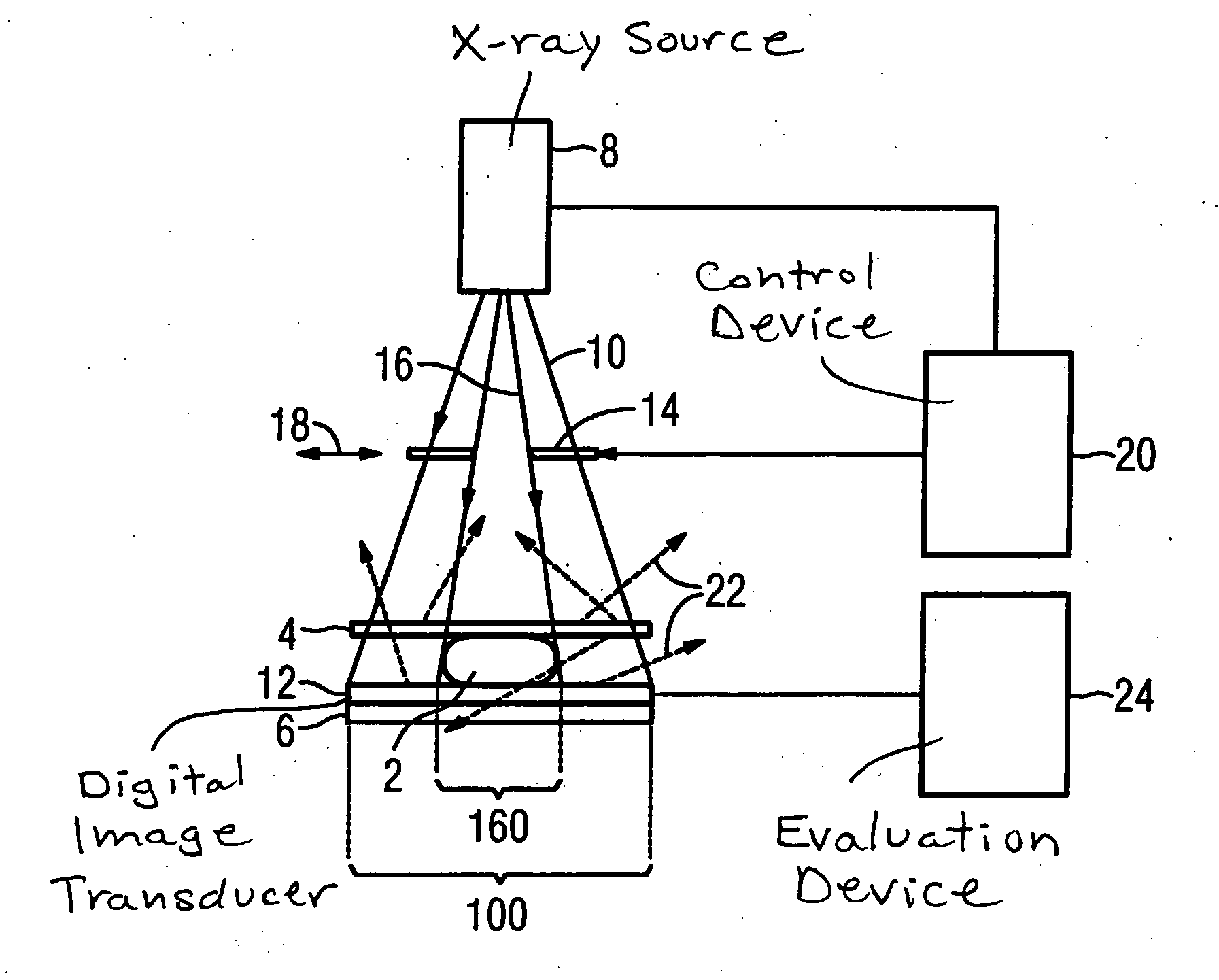
[0014] According to FIG. 1, in an apparatus according to the invention the female breast 2 to be examined is disposed between an upper compression plate 4 and a lower compression plate 6. An x-ray source 8 that generates an approximately conical x-ray beam 10 is located above the upper compression plate 4. The upper compression plate 4 facing the x-ray source 8 is transparent for x-ray radiation. An x-ray receiver is arranged on the lower compression plate 6. Serving as an x-ray receiver is a digital image transducer 12 that converts the incident x-ray quanta into a digital image that can be reproduced, for example, on a monitor.
[0015] A collimator 14 with which the position and cross-section of a partial beam 16 of the x-ray beam 10 impinging on the upper compression plate can be adjusted is disposed in the beam path of the x-ray beam 10. This is symbolically illustrated in the example by the double arrow 18. For this purpose, the collimator 14 can be adjusted by motors. The posit...
PUM
 Abstract
Description
Claims
Application Information