Self-assembling split-fluorescent protein systems

Active Publication Date: 2005-10-06
TRIAD NAT SECURITY LLC
View PDF5 Cites 24 Cited by
  • Summary
  • Abstract
  • Description
  • Claims
  • Application Information

AI Technical Summary

Benefits of technology

[0015] The split-GFP system is very simple, requires no external reagents, provides a sensitive analytical signal directly proportional to the amount of tagged protein, quantifies amounts of proteins typically encountered at the bench in less than 15 minutes, can report either soluble or insoluble protein, and works both in vivo and in vitro. No other existing protein tagging and detection system combines these capabilities. As detailed in the Examples, infra, the split-GFP system has been used to quantify proteins in multiwell plates, and to monitor protein expression and solubility in living Escherichia coli cells.

Problems solved by technology

No other existing protein tagging and detection system combines these capabilities.

Method used


Image

  • Self-assembling split-fluorescent protein systems
  • Self-assembling split-fluorescent protein systems
  • Self-assembling split-fluorescent protein systems

Examples


example 1

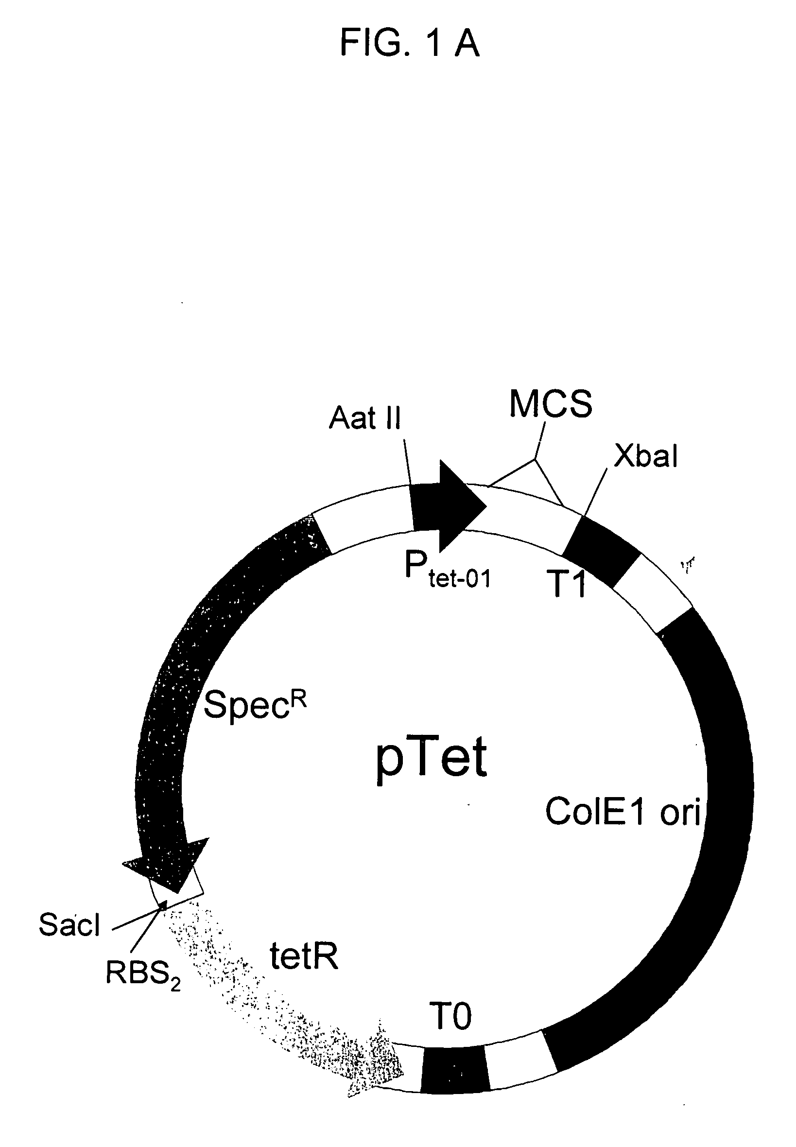
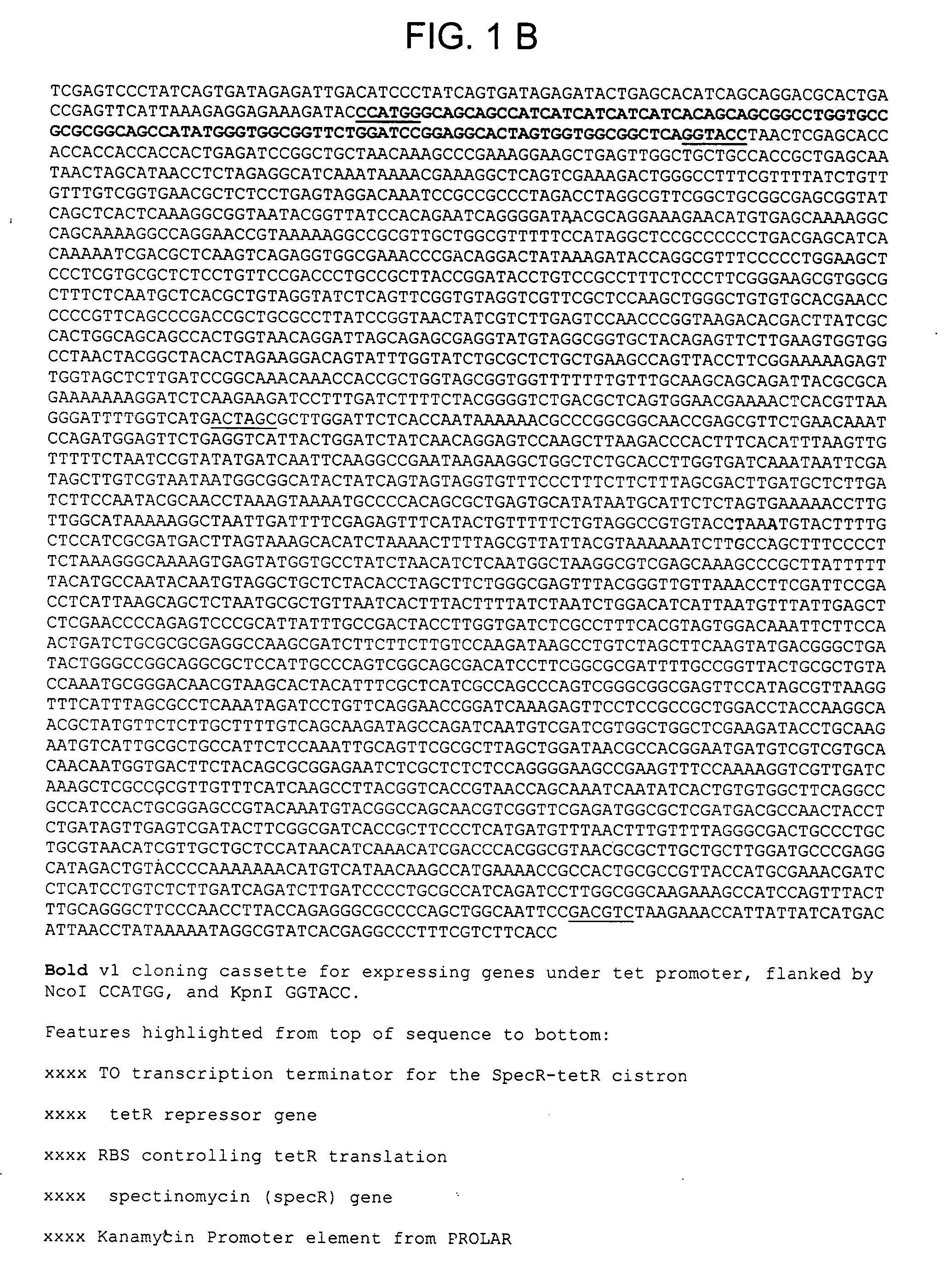
Constructing Plasmid pTET-SpecR

[0141] The commercial tet-promoter PRO Bacterial expression system (Clontech, Palo Alto, Calif.) has the regulatory protein tetR on a second plasmid separate from the expression plasmid, making the creation of large libraries inefficient. To overcome this limitation, we combined the tet promoter which controls the expression of target proteins, and regulatory protein tetR, on a single plasmid containing the tetracycline-inducible promoter tet, the tet promoter regulatory protein tetR, and the selectable antibiotic marker SpecR, which confers resistance to the antibiotic spectinomycin. The ColE1 origin of replication allows this plasmid to co-exist in cells carrying plasmids with a compatible origin such as the p15 origin. This allows one protein, such as a protein tagged with a fragment of GFP, to be expressed from the pTET plasmid, and another protein, such as the complementary GFP assay fragment, to be expressed from a second plasmid, such as a pET ...

example 2

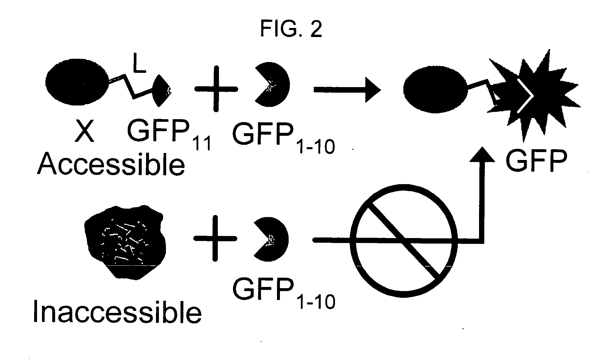
Finding Feasible Pairs of Split GFP

[0150] To achieve the split GFP protein tagging and detection scheme outlined in FIG. 2, we first tested several pairs of fragments from either folding reporter GFP, which bears the mutations F99S, M153T, V163A (Crameri, Whitehorn et al. 1996), F64L, and S65T (Patterson, Knobel et al. 1997), or the exceptionally stable “superfolder” GFP, containing the folding reporter GFP mutations and S30R, Y39N, N105T, Y145F, I171V, and A206V. We separately co-expressed several pairs of GFP fragments on compatible plasmids in E. coli, including amino acids 1-145+145-238, 1-155+156-238, 1-171+171-238, 1-195+196-238, 1-214+214-238. The junction points corresponded to loops or turns between β-strands (Tsien 1998; Baird, Zacharias et al. 1999) (see FIG. 3). Fragment pairs from superfolder GFP consistently gave much brighter colonies than the same pairs from folding reporter GFP. For example, superfolder GFP fragments from split at 156 and 172 were brighter than fra...

example 3

Engineering the GFP Assay Fragment GFP 1-10

[0152] We evolved superfolder GFP 1-10 by DNA shuffling (Stemmer 1994) to improve its solubility and increase its complementation with sulfite reductase-GFP 11. Superfolder GFP 1-10 PCR amplicons were subjected to DNA fragmentation and shuffling using published protocols (Stemmer 1994). The GFP 1-10 cDNA library plasmid was transformed into an E. coli BL21 (DE3) PRO expression strain (Clontech, Palo Alto, Calif.) containing the sulfite reductase-GFP S11 wild type tagged protein on a pPROTET vector (Clontech, Palo Alto, Calif.). The expression library was plated on nitrocellulose membrane using two successive 400-fold dilutions of a 1.0 OD600 nm frozen 20% glycerol / Luria-Bertani (LB) stock. After overnight growth at 37° C., the membrane was transferred to an LB / Agar plate containing 50 μg kanamycin, 35 μg chloramphenicol, and 50 μg spectinomycin per ml of media, plus 1 mM IPTG for 3 h at 37° C., and then moved onto a new plate containing th...


PUM

PropertyMeasurementUnit
Solubility (mass)aaaaaaaaaa
Fluorescenceaaaaaaaaaa

Abstract

The invention provides a protein labeling and detection system based on self-complementing fragments of fluorescent and chromophoric proteins. The system of the invention is exemplified with various combinations of self-complementing fragments derived from Aequorea victoria Green Fluorescent Protein (GFP), which are used to detect and quantify protein solubility in multiple assay formats, both in vitro and in vivo.

Description

RELATED APPLICATIONS [0001] This patent application claims the benefit of the filing date of U.S. Provisional patent application No. 60 / 514,363 under 35 U.S.C. 119(e).STATEMENT AS TO RIGHTS TO INVENTIONS MADE UNDER FEDERALLY SPONSORED RESEARCH OR DEVELOPMENT [0002] This invention was made with government support under grant number DE-FG02-98ER62647 from the United States Department of Energy and Contract No. W-7405-ENG-36 awarded by the United States Department of Energy to The Regents of The University of California. The government has certain rights in this invention.STATEMENT REGARDING COLOR DRAWINGS [0003] This patent or application file contains at least one drawing executed in color. Copies of this patent or patent application publication with color drawings will be provided by the United States Patent and Trademark Office upon request and payment of the necessary fee. BACKGROUND OF THE INVENTION [0004] Obtaining sufficient amounts of soluble, well-folded recombinant proteins ...

Claims


Application Information

Patent Timeline
IPC IPC(8): C07H21/04C07K1/00C07K14/435C12N15/09C12P21/06C12Q1/68G01N33/542G01N33/58
CPCC07K14/43595C07K2319/735C12N15/1058C12N15/1065C12Q1/6897G01N33/542G01N33/582
Inventor WALDO, GEOFFREYCABANTOUS, STEPHANIE
Owner TRIAD NAT SECURITY LLC
Why Eureka
Try Eureka