Automated imaging system for single molecules

Active Publication Date: 2005-10-20
WISCONSIN ALUMNI RES FOUND
View PDF3 Cites 27 Cited by
  • Summary
  • Abstract
  • Description
  • Claims
  • Application Information

AI Technical Summary

Benefits of technology

[0015] This disclosure provides devices and methods for automated collection of images of single molecules and their assemblies, and for preparation of these single molecule images for further processing in external system(s). Both the devices, such as computer systems, and the methods for automated collection and processing provide for an overall robust imaging operation. The systems may include one or more of the following features, or may utilize all of them. Focus is advantageously automatically maintained during image collection. During the image processing step, the uneven illumination of fluorescence microscopy may be substantially reduced or eliminated by a flattening process. Offset of overlap may be determined between images and adjusted. Overly-exposed (bleached) region of overlap on one image

Problems solved by technology

It is however a challenge to devise practical ways to extract meaningful data from large datasets of molecular images.
However, proper statistical analysis, necessary for the accurate assessment of physical, chemical and biochemical quantities, requires larger datasets, and it has remained intrinsically difficult to generate these datasets in single molecule studies due to image analysis and file management issues.
Practical issues in pixel-based autofocusing for machine vision.
This may be due to the fact that varying types of tissues (cells, DNA, etc.) each present their own set of automatic focusing cha

Method used


Image

  • Automated imaging system for single molecules
  • Automated imaging system for single molecules
  • Automated imaging system for single molecules

Examples


example 1

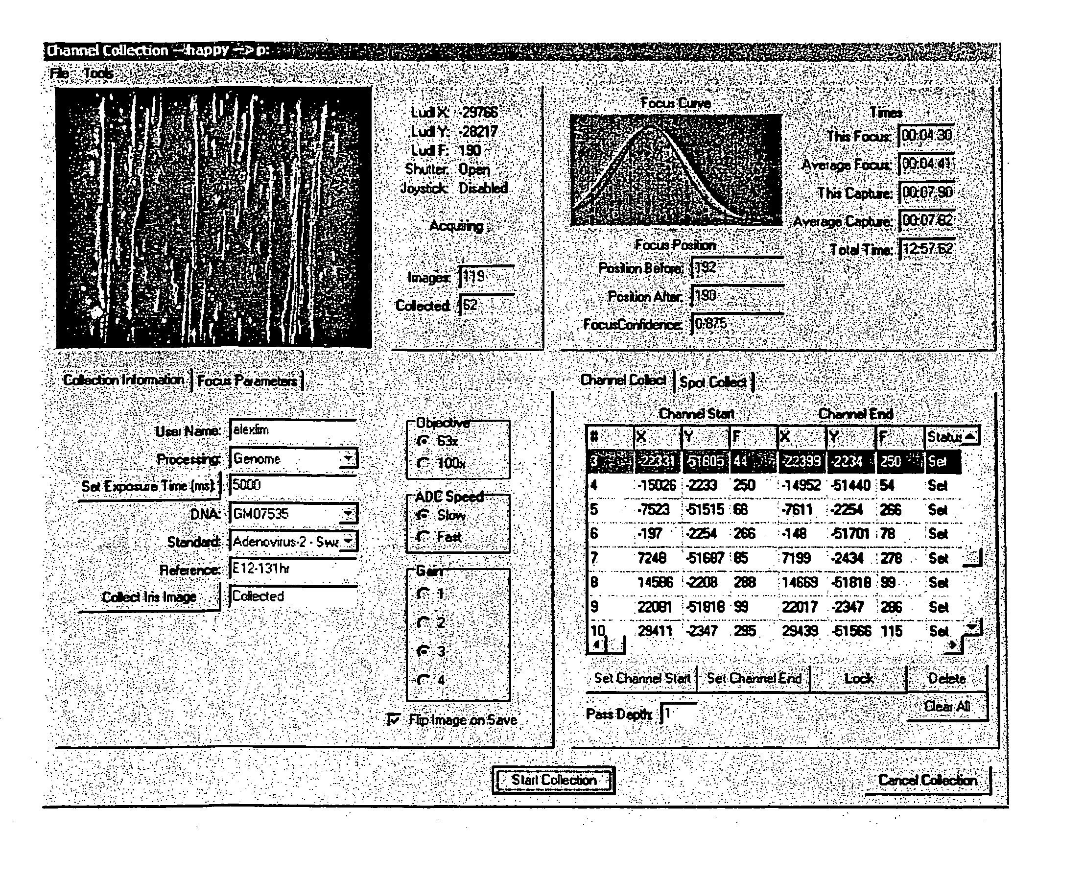
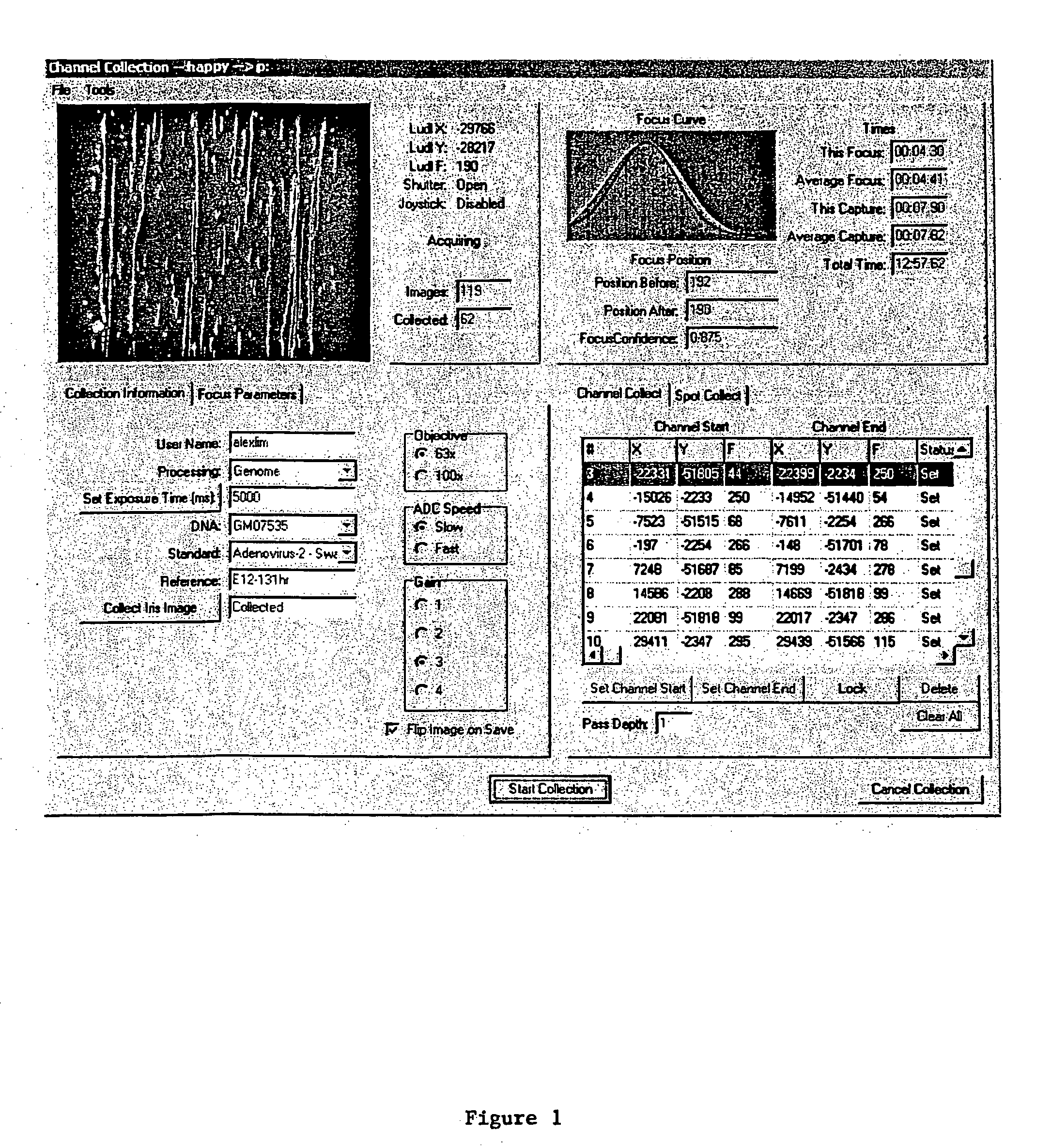
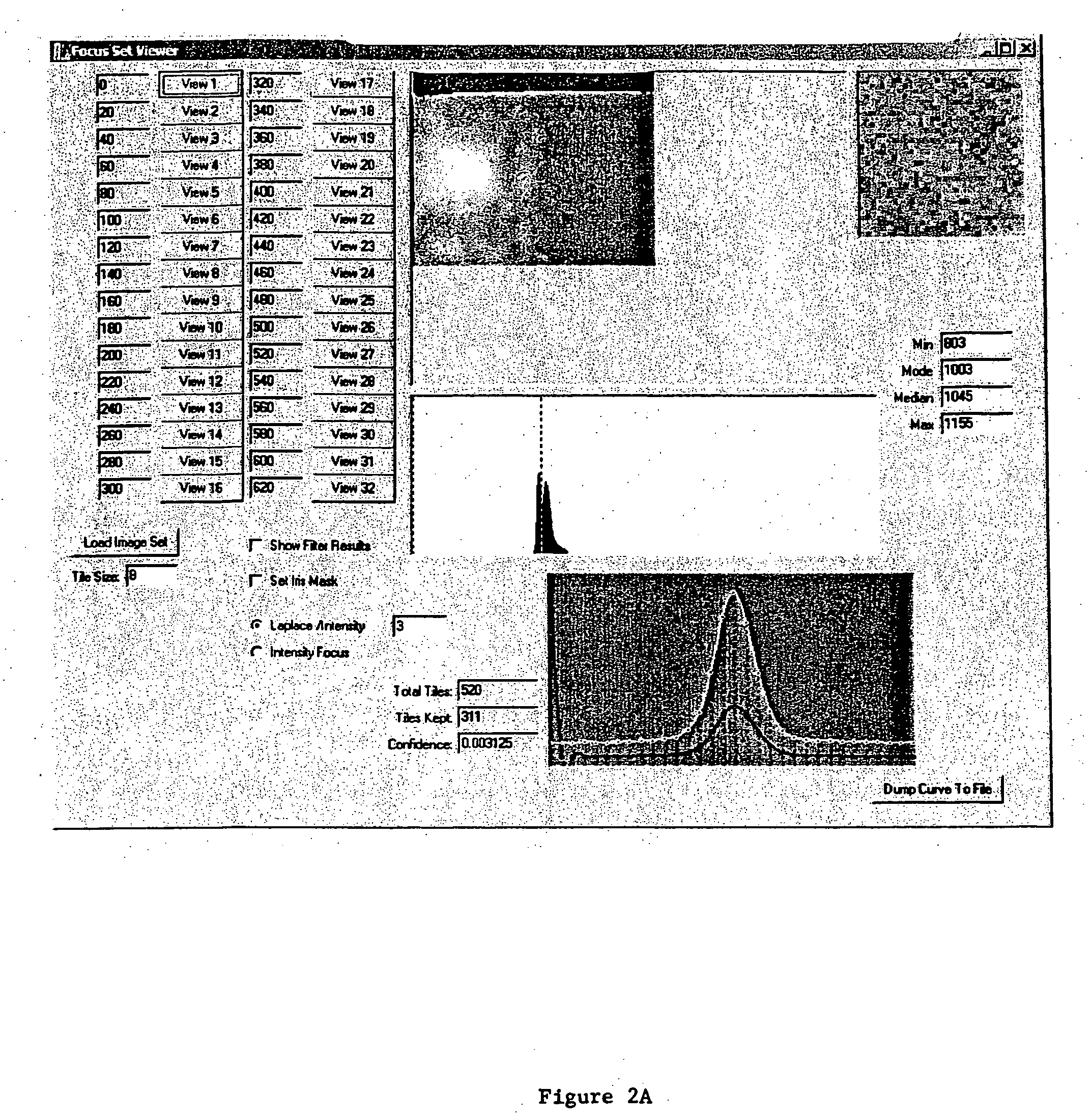
The Automated Focus Component Routine

[0111] The automated focus component routine is written to work with an interface to a CCD camera. Since more than one type of CCD camera may be used, C++ was used to develop an abstract class to encompass a variety of camera classes. During the setup phase the type of camera is queried from the object to determine both allowed binning values and optimal exposure times. The automated focus component object also assumes the existence of a translatable Z axis (motorized objective column), in various embodiments with LUDL access. Some classes that the automated focus component object uses are not documented here (SmoothingSplines for example) but are well understood in the art. The specifics of the LUDL stage controller and varying CCD camera drivers are also accessed through classes (as mentioned supra) and provide for a clearer and more flexible solution.

example 3

Code for the Overlap Program of the System and Method Disclosed

[0112] The command line used to run the program for sub-images S1 and S2, and CCF region shown in FIGS. 3A-C and discussed below is: [0113] overlap raw1-2212017.omi raw1-2212016.omi-13 810 [0114] where (−13, 810) is the initial overlap estimate, meaning (0,0) in S1 is at (−13, 810) in S2. The output is: [0115] raw1-2212017.omi raw1-2212016.omi −35 774 0 −35.22 774.42 0.700 0.361

which indicates that the true offset is (−35, 774) and overlap is good (with zero status). The sub-pixel alignment from fitting the two-dimensional parabola is (−35.22, 774.42) with a correlation peak of 0.700 and a total variance of 0.361. Note that (−13, 810) minus (−35, 774) equals (22, 36) which is the (sub-)offset of the correlation peak from the center of the cross-hairs.


PUM


Abstract

There is provided a high throughput automated single molecule image collection and processing system that requires minimal initial user input. The unique features embodied in the present disclosure allow automated collection and initial processing of optical images of single molecules and their assemblies. Correct focus may be automatically maintained while images are collected. Uneven illumination in fluorescence microscopy is accounted for, and an overall robust imaging operation is provided yielding individual images prepared for further processing in external systems. Embodiments described herein are useful in studies of any macromolecules such as DNA, RNA, peptides and proteins. The automated image collection and processing system and method of same may be implemented and deployed over a computer network, and may be ergonomically optimized to facilitate user interaction.

Description

[0001] The work described in this disclosure was conducted with United States Government support, grant number DE-FG02-99ER62830, awarded by the Department of Energy. The United States Government has certain rights in the invention(s) of this disclosure.BACKGROUND OF THE DISCLOSURE [0002] 1. Field of the Disclosure [0003] The present disclosure relates to a high throughput automated single molecule image collection and processing system that requires minimal or limited initial user input. Optical images of single molecules and fragments elongated and fixed within microfluidic channels can be automatically collected, maintaining correct focus, and the images prepared for further data processing. A computer-based analysis can be performed on each image thereby obviating the problem of uneven illumination in fluorescence microscopy, and providing an overall robust imaging operation. Embodiments described herein are thus useful in studies of any macromolecules such as DNA, RNA and prote...

Claims


Application Information

Patent Timeline
IPC IPC(8): G06F19/00G06K9/00
CPCG06K9/00134G06V20/693
Inventor SCHWARTZ, DAVID CHARLESRUNNHEIM, RODNEYFORREST, DANIEL
Owner WISCONSIN ALUMNI RES FOUND
Why Eureka
Try Eureka