Aqueous outflow enhancement with vasodilated aqueous cavity

a vasodilated aqueous cavity and enhancement technology, applied in the field of reducing intraocular pressure within the animal eye, can solve the problems of large surgical skill, substantial trauma to the eye, and low success rate, and achieve the effects of enhancing aqueous outflow, reducing intraocular pressure, and being safer and less expensiv

Inactive Publication Date: 2005-11-10
GLAUKOS CORP
View PDF99 Cites 185 Cited by
  • Summary
  • Abstract
  • Description
  • Claims
  • Application Information

AI Technical Summary

Benefits of technology

[0019] All of the above embodiments and variations thereof have numerous disadvantages and moderate success rates. They involve substantial trauma to the eye and require great surgical skill in creating a hole through the full thickness of the sclera into the subconjunctival space. The procedures are generally performed in an operating room and have a prolonged recovery time for vision.
[0023] What is needed, therefore, is an extended, site specific treatment method for glaucoma that is faster, safer, and less expensive than currently available modalities. It is one object of the invention to provide a drug-eluting trabecular stent with vasodilating capability enabling enhanced aqueous outflow and lowered intraocular pressure.
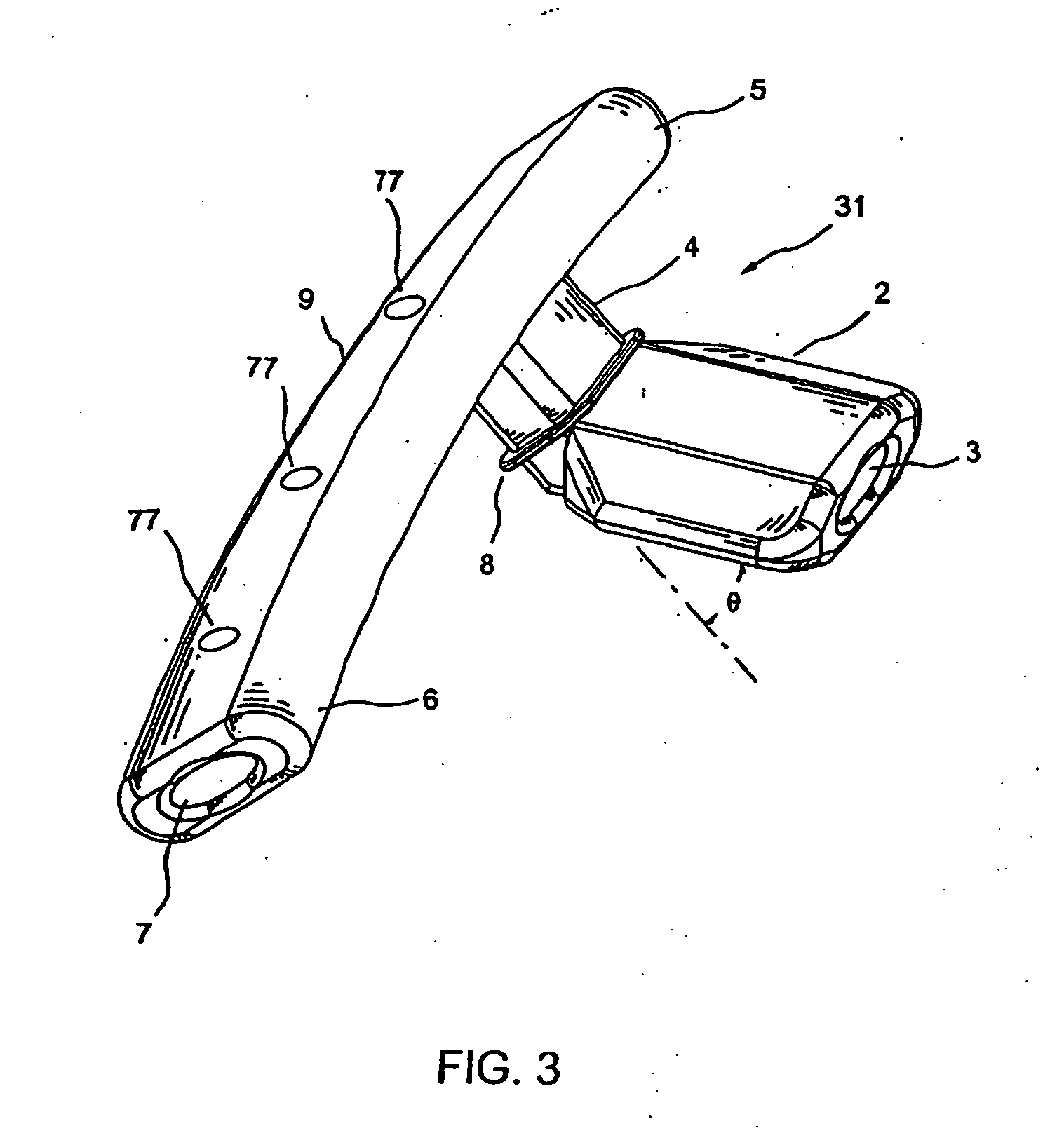
[0024] A device and method are provided for improved treatment of intraocular pressure due to glaucoma. A trabecular stenting device is adapted for implantation within a trabecular meshwork of an eye such that aqueous humor flows controllably from an anterior chamber of the eye to Schlemm's canal, bypassing the trabecular meshwork. In one embodiment, the trabecular stenting device comprises a quantity of pharmaceuticals effective in treating glaucoma, which are controllably released from the device into tissue of Schlemm's canal and / or downstream collector channels. Depending upon the specific treatment contemplated, pharmaceuticals may be utilized in conjunction with the trabecular stenting device such that aqueous flow either increases or decreases as desired. Placement of the trabecular stenting device within the eye and incorporation, and eventual release, of a proven pharmaceutical glaucoma therapy will reduce, inhibit or slow the effects of glaucoma.

Problems solved by technology

All of the above embodiments and variations thereof have numerous disadvantages and moderate success rates.
They involve substantial trauma to the eye and require great surgical skill in creating a hole through the full thickness of the sclera into the subconjunctival space.
However, modifying existing filtering surgery techniques in any profound way to increase their effectiveness appears to have reached a dead end.

Method used


Image

  • Aqueous outflow enhancement with vasodilated aqueous cavity
  • Aqueous outflow enhancement with vasodilated aqueous cavity
  • Aqueous outflow enhancement with vasodilated aqueous cavity

Examples


Embodiment Construction

[0063] The preferred embodiments of the present invention described below relate particularly to surgical and therapeutic treatment of glaucoma through reduction of intraocular pressure. While the description sets forth various embodiment specific details, it will be appreciated that the description is illustrative only and should not to be construed in any way as limiting the invention. Furthermore, various applications of the invention, and modifications thereto, which may occur to those who are skilled in the art, are also encompassed by the general concepts described below.

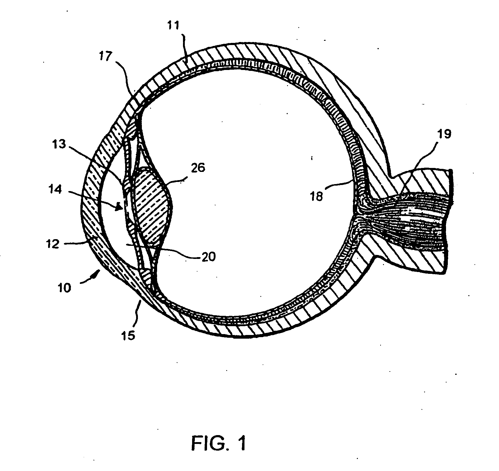
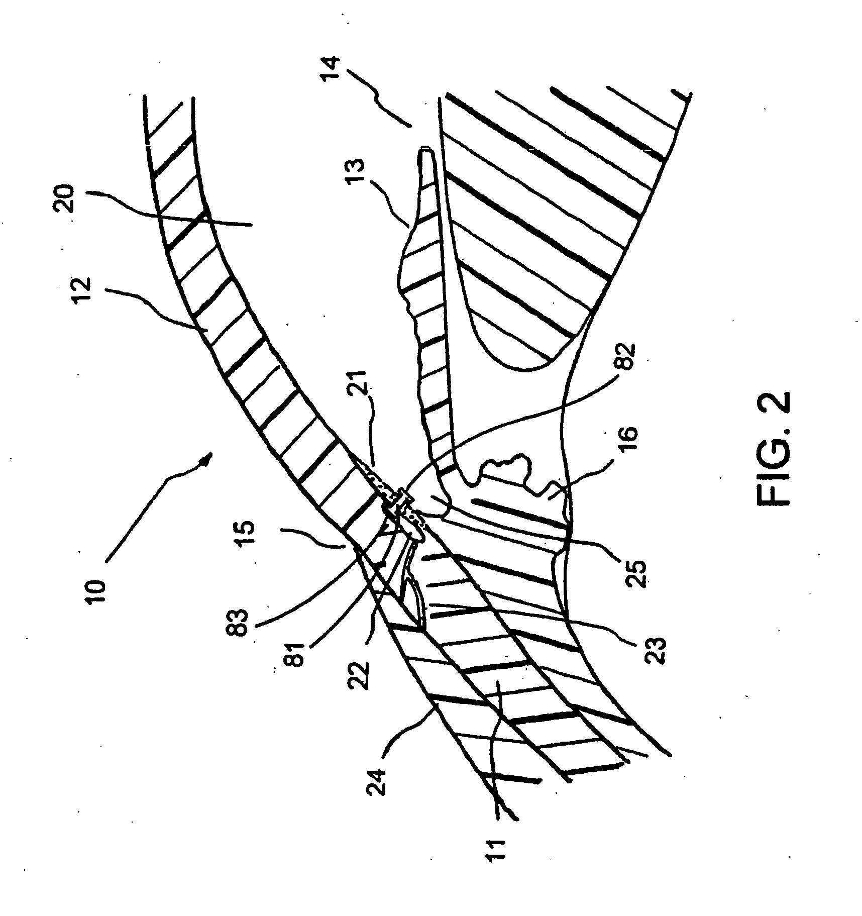
[0064]FIG. 1 is a cross-sectional view of an eye 10, while FIG. 2 is a close-up view showing the relative anatomical locations of a trabecular meshwork 21, an anterior chamber 20, and a Schlemm's canal 22. A sclera 11 is a thick collagenous tissue which covers the entire eye 10 except a portion which is covered by a cornea 12. The cornea 12 is a thin transparent tissue that focuses and transmits light into th...


PUM

PropertyMeasurementUnit
angleaaaaaaaaaa
angleaaaaaaaaaa
lengthaaaaaaaaaa

Abstract

A method for enhancing aqueous outflow and thereby lowering intraocular pressure is disclosed. The method comprises vasodilating aqueous veins by dilating or relaxing the smooth muscle of an aqueous cavity. In one embodiment, the step of dilating or relaxing the smooth muscle of the aqueous cavity is accomplished by slowly releasing loaded smooth muscle relaxing drug at an effective dose over time. In another embodiment, the step of dilating the smooth muscle of the aqueous cavity is accomplished by introducing a smooth muscle drug through an implant.

Description

RELATED APPLICATIONS [0001] This application claims priority from U.S. Provisional Application No. 60 / 540,521, entitled “Aqueous Outflow Enhancement with Vasodilated Aqueous Cavity,” filed Jan. 30, 2004, the entirety of which is hereby incorporated by reference.BACKGROUND OF THE INVENTION [0002] 1. Field of the Invention [0003] This invention relates to reducing intraocular pressure within the animal eye. More particularly, this invention relates to a treatment of glaucoma wherein aqueous humor is permitted to flow out of an anterior chamber of the eye through a surgically implanted pathway. Furthermore, this invention relates to a direct delivery of pharmaceuticals to ocular tissue in the eye. [0004] 2. Description of the Related Art [0005] The human eye is a specialized sensory organ capable of light reception and able to receive visual images. The trabecular meshwork serves as a drainage channel and is located in the anterior chamber angle formed between the iris and the cornea. ...

Claims


Application Information

Patent Timeline
Patent Type & Authority Applications(United States)
IPC IPC(8): A61K31/519
CPCA61F9/00781A61K31/519A61K9/0051A61K9/0048
Inventor TU, HOSHENGHAFFNER, DAVID
Owner GLAUKOS CORP
Why Eureka
Try Eureka