
Medical device having radio-opacification and barrier layers

a barrier layer and radio-opacification technology, applied in the field of invivo stents, can solve the problems of myocardial infarction, abrupt closure and restnosis of angioplasty procedures, and achieve the effect of reducing conta

Inactive Publication Date: 2005-11-10
BOSTON SCI SCIMED INC
View PDF19 Cites 7 Cited by
  • Summary
  • Abstract
  • Description
  • Claims
  • Application Information

AI Technical Summary

Benefits of technology

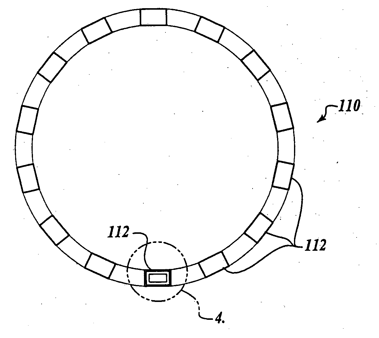
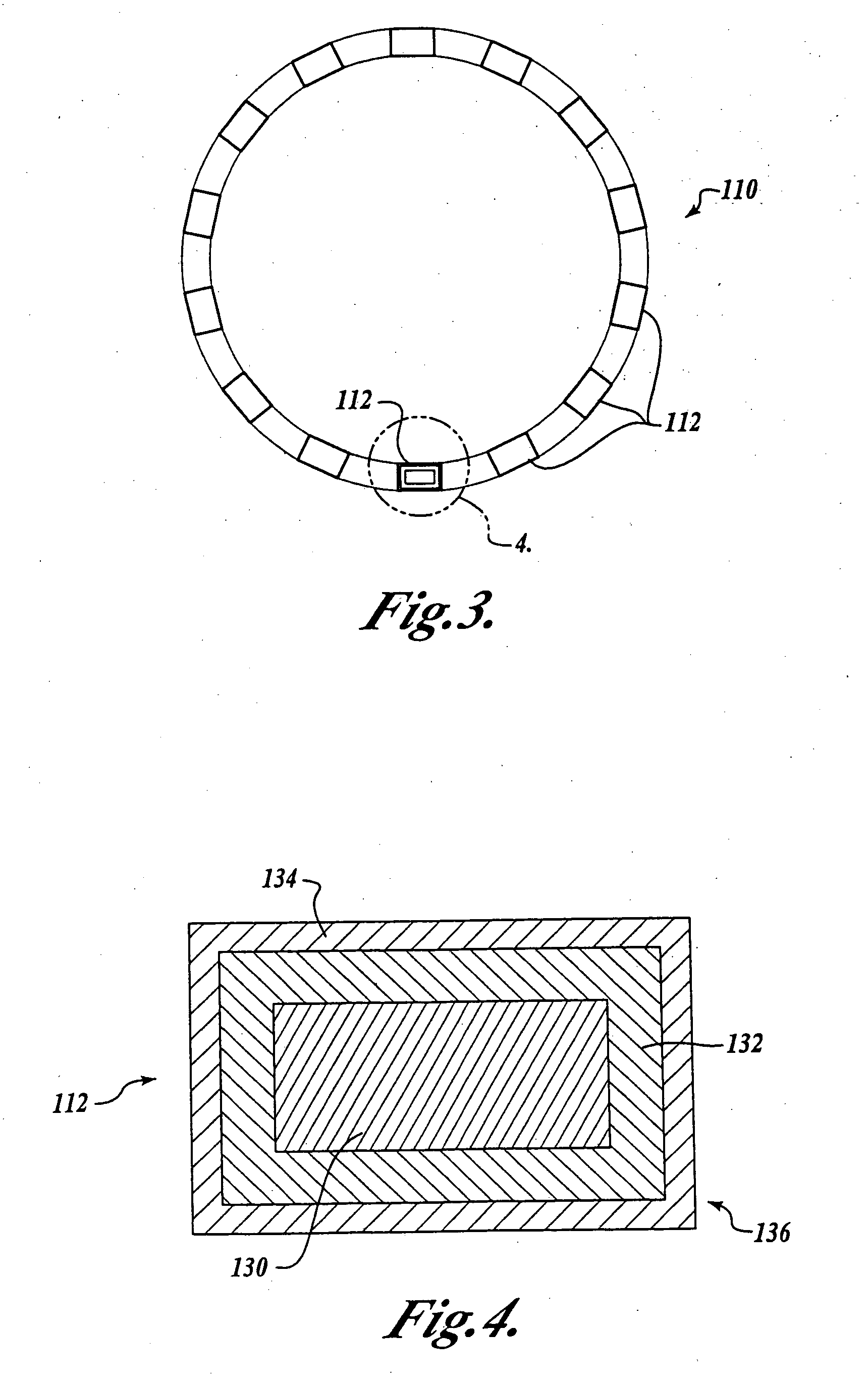
[0009] In accordance with a first aspect of the present invention, a laminate structure is provided for making a medical device. The laminate structure comprises a core having an outer surface and a first layer secured onto a portion of the outer surface of the core. The first layer has an outer surface and is radio-opaque. A second bio-compatible layer is secured onto at least a portion of the outer surface of the first layer to reduce contact between the first layer and blood and / or tissue in a vessel.

Problems solved by technology

While PTA and PTCA have gained wide acceptance, these angioplasty procedures suffer from two major problems: abrupt closure and restenosis.
Abrupt closure refers to rapid re-occlusion of the vessel immediately after or within hours of the initial treatment, and often can result in myocardial infarction if blood flow is not restored in a timely manner.
However, the preferred structural material, stainless steel, used in stents is not highly radio-opaque.

Method used


Image

  • Medical device having radio-opacification and barrier layers
  • Medical device having radio-opacification and barrier layers
  • Medical device having radio-opacification and barrier layers

Examples


Embodiment Construction

[0022] While, as will be better understood from the following description, the present invention was developed for coronary stents and, thus, is expected to find its primary use with such coronary stents, it is to be understood that the invention can be used with other medical devices such as vena cava filters, aneurysm coils or other implantable devices that require the ability to be visualized in-vivo and to have a bio-compatible barrier layer. Thus, it is to be understood that the disclosed embodiment is only by way of example and should not be construed as limiting.

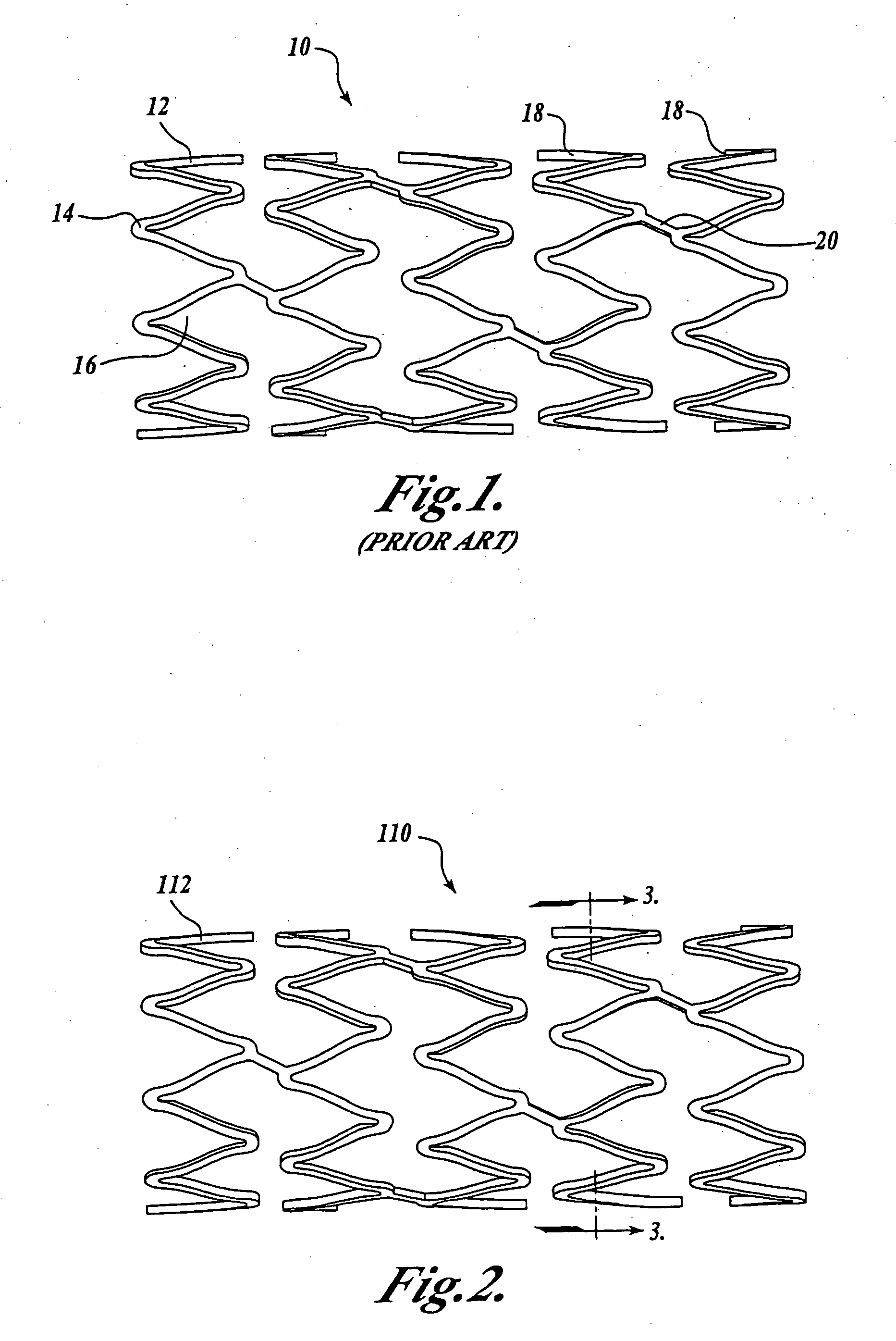
[0023] Prior to describing an illustrative embodiment of the invention, a brief discussion of the structure of one type of medial device is set forth. In this regard, attention is directed to FIG. 1, which illustrates a conventional medical device known in the art as a coronary stent 10. The coronary stent 10 is deployed in-vivo at a stenosed vessel following a PTCA procedure. The stent 10 is deployed from a delivery...


PUM


Abstract

A medical device such as a coronary stent is provided that can be visualized in-vivo while further aiding in the prevention of restenosis. The medical device comprises a core having a first layer disposed thereon. The first layer is made from a material that is radio-opaque so that the medical device may be visualized in-vivo. An outer layer is disposed onto and surrounds at least a portion of the first layer to provide a barrier layer between the radio-opaque inner layer and blood and / or tissue disposed within the patient's vessel. The outer surface of the outer layer may include a textured surface of micro-pores, grooves, cross-hatched lines to receive a therapeutic agent. Drugs and treatments which utilize anti-thombogenic agents, and anti-proliferation agents may be readily deployed from the textured outer surface of the outer layer of the medical device.

Description

FIELD OF THE INVENTION [0001] The present invention relates generally to devices for preventing vascular diseases, and more specifically to in-vivo stents used in medical procedures. BACKGROUND OF THE INVENTION [0002] As an alternative to vascular surgery, percutaneous transluminal angioplasty (PTA) and percutaneous transluminal coronary angioplasty (PTCA) procedures are being widely used for treating stenotic atherosclerotic regions of a patient's vasculature to restore adequate blood flow. Catheters having an expansible distal end, typically in the form of an inflatable balloon, are positioned in a vessel, such as a coronary artery, at a stenotic site. The expansible end is then expanded to dilate the vessel in order to restore adequate blood flow to regions beyond the stenosis. While PTA and PTCA have gained wide acceptance, these angioplasty procedures suffer from two major problems: abrupt closure and restenosis. [0003] Abrupt closure refers to rapid re-occlusion of the vessel ...

Claims


Application Information

Patent Timeline
IPC IPC(8): A61F2/915A61L31/00A61L31/12A61L31/18
CPCA61L31/18A61L31/121
Inventor CHANDRESEKARA, VERIVADA CHANDRUKVEEN, GRAIG
Owner BOSTON SCI SCIMED INC
Learn More