Four-dimensional imaging of periodically moving objects via post-acquisition synchronization of nongated slice-sequences

a four-dimensional, non-gated technology, applied in the field of cardiovascular medical imaging, can solve the problems of difficult reliable triggering signals to gate the acquisition, cumbersome acquisition, and slow confocal microscopes at successive depths, and achieve the effect of rapid execution

Inactive Publication Date: 2005-11-24
CALIFORNIA INST OF TECH
View PDF10 Cites 79 Cited by
  • Summary
  • Abstract
  • Description
  • Claims
  • Application Information

AI Technical Summary

Benefits of technology

[0015] To solve some of the above requirements, our synchronization algorithms rely on the wavelet transform for robustness and rapid execution. The use of wavelet properties to implem

Problems solved by technology

Despite recent technological breakthroughs in two dimensional dynamic imaging, confocal microscopes remain quite slow at capturing optical sections at successive depths.
For smaller organisms, such as the zebrafish em

Method used


Image

  • Four-dimensional imaging of periodically moving objects via post-acquisition synchronization of nongated slice-sequences
  • Four-dimensional imaging of periodically moving objects via post-acquisition synchronization of nongated slice-sequences
  • Four-dimensional imaging of periodically moving objects via post-acquisition synchronization of nongated slice-sequences

Examples


Embodiment Construction

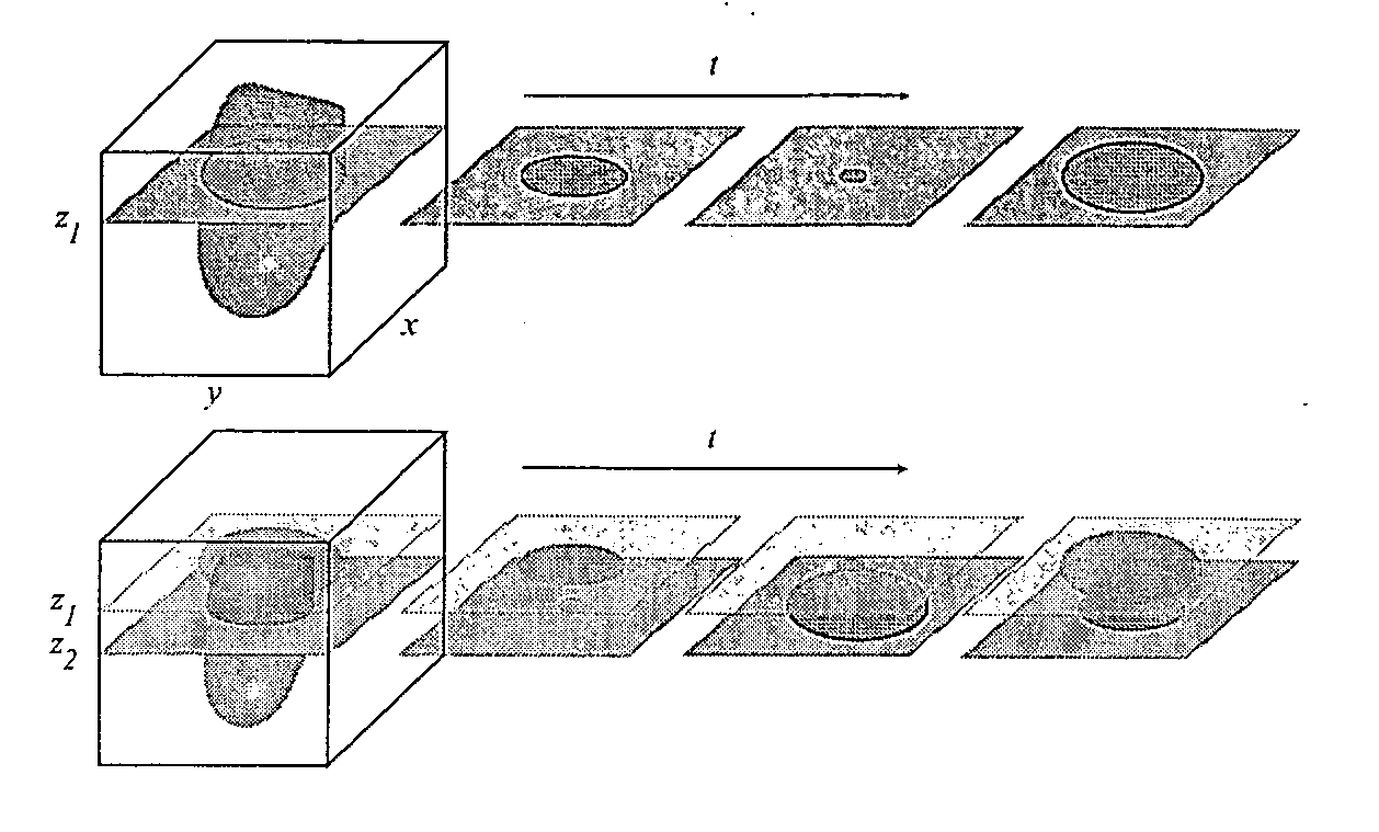
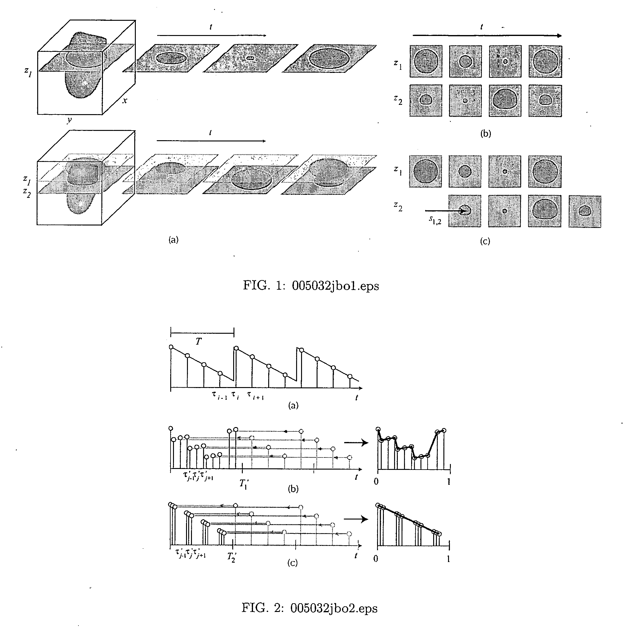
[0027] Fluctuations in fluorescence intensities produced by regular cycles can be used to register and segment images. Any regular sequence can be used to register four-dimensional data or can be used to identify individual fluorescence particles or cells within a preparation, if images are collected with a high enough frame rate. The availability of fast scanning confocal microscopes, for instance, make it possible to collect optical sectioned data in fast temporal sequences.

[0028] For instance, the regular beat of the heart can be used to align time-lapse images taken at different focal planes. This makes it possible to collect rapid time sequences of three dimensional data. Since images taken sequentially on the same plane can be acquired more rapidly that images collected on different optical planes, time sequences can be acquired at separate optical planes and then can be registered with respect to the temporal alignment using the regular rhythm of the cardiac cycle.

[0029] Re...


PUM


Abstract

When the studied motion is periodic, such as for a beating heart, it is possible to acquire successive sets of two dimensional plus time data slice-sequences at increasing depths over at least one time period which are later rearranged to recover a three dimensional time sequence. Since gating signals are either unavailable or cumbersome to acquire in microscopic organisms, the invention is a method for reconstructing volumes based solely on the information contained in the image sequences. The central part of the algorithm is a least-squares minimization of an objective criterion that depends on the similarity between the data from neighboring depths. Owing to a wavelet-based multiresolution approach, the method is robust to common confocal microscopy artifacts. The method is validated on both simulated data and in-vivo measurements

Description

RELATED APPLICATIONS [0001] The present application is related to U.S. Provisional Patent Application, Ser. No. 60 / 568,574, filed on May 5, 2004, which is incorporated herein by reference and to which priority is claimed pursuant to 35 USC 119.BACKGROUND OF THE INVENTION [0002] 1. Field of the Invention [0003] The invention relates to the field of confocal laser scanning microscopy (CLSM) and in particular cardiac medical imaging. [0004] 2. Description of the Prior Art [0005] Confocal laser scanning microscopy (CLSM) has emerged as a popular method for high resolution imaging of fluorescent labels, particularly in thick or scattering samples. By placing a pinhole in the conjugate optical plane, before the detector, out-of-focus light from above and below the focal plane is rejected from the image, enhancing the axial resolution. By collecting images from defined optical slices at successive depths, the three-dimensional arrangement of fluorescently-labeled structures can be derived....

Claims


Application Information

Patent Timeline
IPC IPC(8): G06K9/00
CPCG06K9/00134G06V20/693
Inventor DICKINSON, MARYFAROUHAR, ARIANFRASER, SCOTT E.GHARIB, MORTEZALIEBLING, MICHAEL
Owner CALIFORNIA INST OF TECH
Why Eureka
Try Eureka