
Electrosurgical ablator with integrated aspirator lumen and method of making same

a technology of ablator and lumen, applied in the field of electrosurgical devices, can solve the problems that the upper active edge of the tissue is not able to prevent gas bubbles from passing into the lumen, and achieve the effect of reducing the plugging

Active Publication Date: 2005-12-15
LINVATEC
View PDF15 Cites 43 Cited by
  • Summary
  • Abstract
  • Description
  • Claims
  • Application Information

AI Technical Summary

Benefits of technology

[0016] The present invention overcomes the disadvantages of the electrosurgical devices in the prior art by providing an electrode that reduces plugging. In an exemplary embodiment, the electrosurgical instrument includes a handle with a probe extending from the handle. The probe can be inserted into a patient during an arthroscopic procedure to ablate tissue.
[0021] The present invention advantageously prevents tissue fragments from plugging the aspirating lumen of the electrosurgical device. The continuous flow of fluids and gasses through the aspirating lumen greatly increases the ability of the physician to complete a procedure without interruption. The surgeon's clear field of vision provided by using the electrosurgical device of the present invention helps prevent errors, increases the speed with which the surgeon can complete the procedure and thus reduces the overall expense of the surgical procedure.
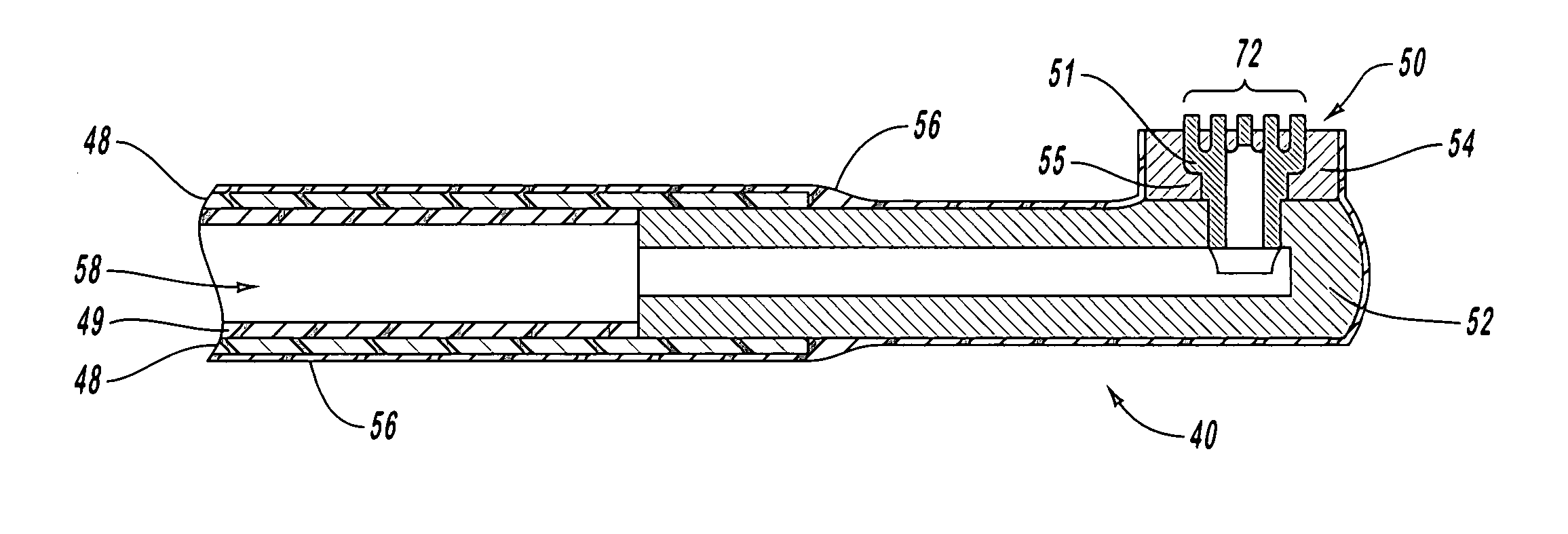
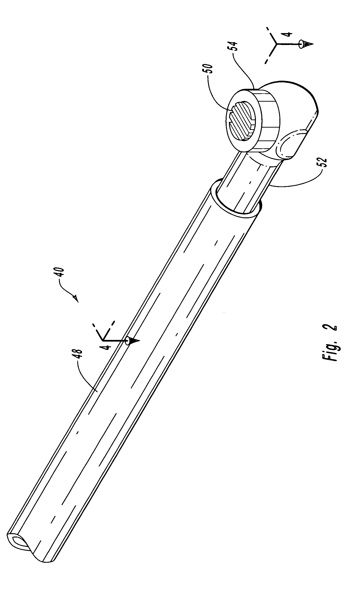
[0022] The method of manufacturing the electrosurgical device of the present invention provides significant advantages over the prior art. In one embodiment of the present invention, the electrode is made from a single piece of electrically conductive material. The lumen is made by back drilling a bore into the piece of material. The bore terminates proximal to the distal end. A series of grooves are then cut into the distal end. The grooves are cut deep enough to reach the lumen, thereby creating openings into the lumen. The grooves also create ribs of material. The upper edges of the ribs form active edges where the electrode arcs, thereby generating the cutting potential of the electrode.
[0023] Creating an electrode in this manner can significantly reduce the cost of manufacturing. Back drilling the bore and cutting the grooves are relatively simple and economical manufacturing procedures, yet they can produce an electrode with extensive amounts of active edges for ablating tissue.
[0024] The probe of an electrosurgical device must be very small for it to be inserted into a patient through the portals, as discussed above. Therefore, creating electrodes with a large active surface area can be particularly challenging. The problems accompanying size restriction are further compounded by adding a sizable lumen to the probe for aspirating gasses and debris. The manufacturing techniques of the present invention optimize lumen size and ablation potential.
[0025] The active area of the electrode is maximized by placing the lumen blow the active area. The placement of the lumen allows for a greater ablation surface, yet the configuration of the ablation surfaces helps prevent clogging of the lumen as compared with prior art devices.

Problems solved by technology

Furthermore, tissue stuck on the upper active edges typically does not prevent gas bubbles from passing into the lumen because of the many alternative paths to the lumen through the upper edges.

Method used


Image

  • Electrosurgical ablator with integrated aspirator lumen and method of making same
  • Electrosurgical ablator with integrated aspirator lumen and method of making same
  • Electrosurgical ablator with integrated aspirator lumen and method of making same

Examples


Embodiment Construction

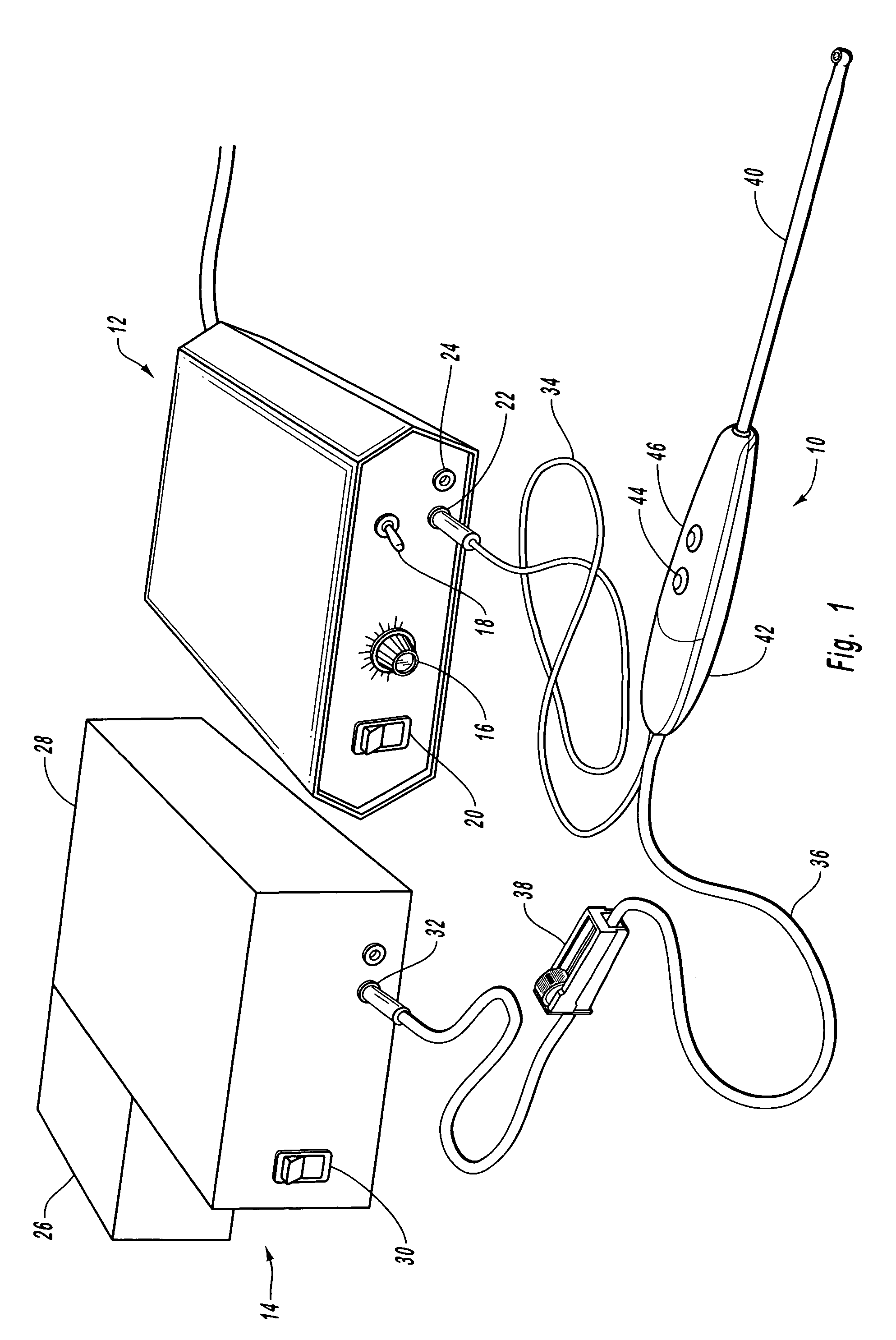
[0034] Embodiments of the present invention relate to electrosurgical systems for ablating tissue in an electrosurgical procedure. FIG. 1 shows an exemplary electrosurgical system which includes an electrosurgical instrument 10 connected to an electrosurgical generator 12 and an aspirator 14.

[0035] In an exemplary embodiment, electrosurgical generator 12 is configured to generate radio frequency (“RF”) wave forms for a monopolar instrument such as electrosurgical instrument 10. Generator 12 can generate energy useful for ablating tissue and / or coagulating tissue. In one embodiment, generator 12 includes standard components, such as dial 16 for controlling the frequency and / or amplitude of the RF energy, a switch 18 for changing the type of waveform generated, a switch 20 for turning the generator on and off, and an electrical port 22 for connecting the electrosurgical instrument 10. Generator 12 also includes port 24 for connecting an electrical ground. It will be appreciated that ...


PUM


Abstract

Electrosurgical devices for ablating tissue and methods of making the same are disclosed. The electrosurgical devices include an electrode with a lumen for aspirating gasses and debris produced during surgery. The electrode also includes a plurality of upper active edges for ablating tissue in an electrosurgical procedure. The upper active electrodes are spaced apart to form a filter that filters out large tissue particles from being aspirated into the lumen. The upper active edges ablate the large particles to form particles that can pass into the lumen. A lower active edge is formed at the terminal end of the lumen for ablating tissue being aspirated into the lumen. The lower active edge prevent tissue particles from occluding the opening to the lumen.

Description

BACKGROUND OF THE INVENTION [0001] 1. The Field of the Invention [0002] The present invention relates to electrosurgical devices for ablating tissue in an arthroscopic procedure. More specifically, the present invention relates to electrosurgical devices with an electrode that defines a lumen for aspirating gasses and debris. [0003] 2. The Relevant Technology [0004] An arthroscope is an instrument used to look directly into a surgical site. Typically, the arthroscope utilizes a magnifying lens and coated glass fibers that beam an intense, cool light into the surgical site. A camera attached to the arthroscope allows the surgeon to view the surgical site on a monitor in the operating room. With the arthroscope, the surgeon can look directly into a surgical site, such as a knee or shoulder, to diagnose injury and decide on the best treatment. While viewing the surgical site with the arthroscope, the surgeon can repair an injury using a separate surgical instrument. [0005] The ability ...

Claims


Application Information

Patent Timeline
Patent Type & Authority Applications(United States)
IPC IPC(8): A61B18/14
CPCA61B2218/007A61B18/148
Inventor DECESARE, MICHAELWEST, HUGH S. JR.
Owner LINVATEC
Learn More