Antibodies against Clostridium difficile toxins and uses thereof

a technology of clostridium difficile and antibodies, which is applied in the field of antibodies against clostridium difficile toxins, can solve the problems of complex treatment of c. difficile, cell death, depolymerization of actin filaments, etc., and achieve the effect of high affinity antibodies

Active Publication Date: 2005-12-29
MASSACHUSETTS UNIV OF +2
View PDF12 Cites 53 Cited by
  • Summary
  • Abstract
  • Description
  • Claims
  • Application Information

AI Technical Summary

Benefits of technology

[0010] This invention is based, in part, on the discovery that administration of antibodies against C. difficile toxin A to a subject can protect the subject from relapse of C. difficile-mediated disease in vivo. Administration of antibodies to one or both of toxin A and toxin B can prevent primary C. difficile-mediated disease. High affi...

Problems solved by technology

Modification of Rho proteins by the toxins inactivates them, leading to depolymerization of actin filaments and ...

Method used


Image

  • Antibodies against Clostridium difficile toxins and uses thereof
  • Antibodies against Clostridium difficile toxins and uses thereof
  • Antibodies against Clostridium difficile toxins and uses thereof

Examples


example 1

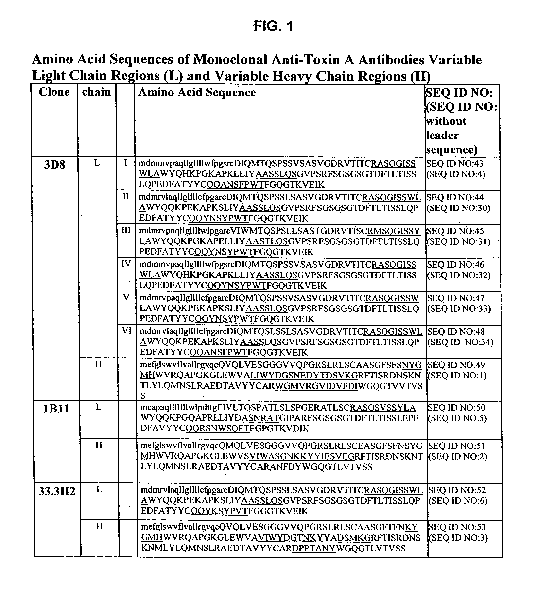
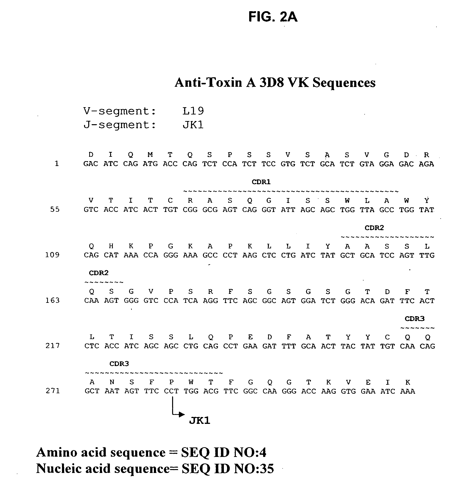
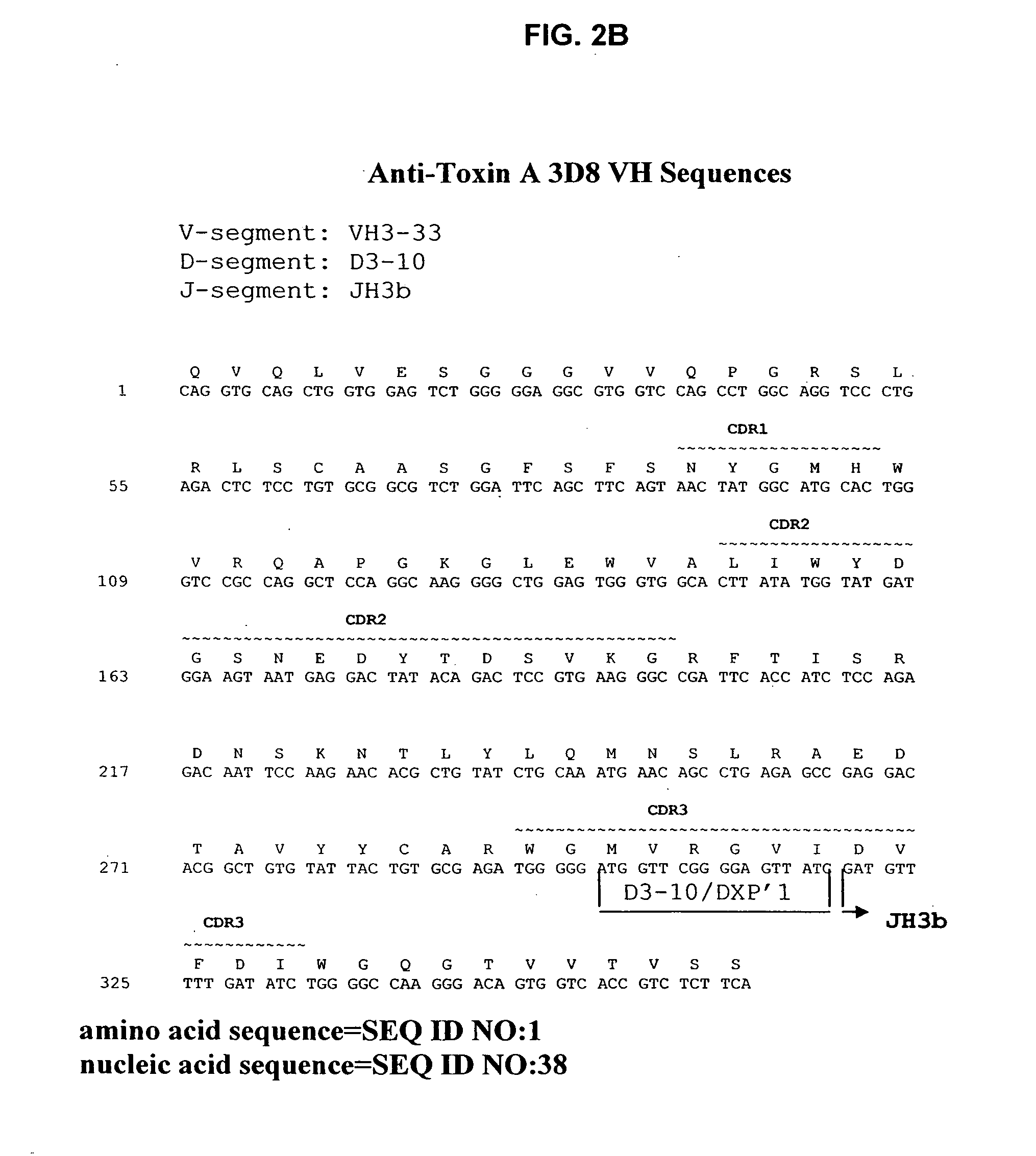
Generation of Anti-Toxin A Monoclonal Antibodies

[0208]C. difficile toxin A was obtained either from Techlab, Inc. (Blacksburg, Va.), or by recombinant production. The toxin was purified and inactivated prior to immunization. Inactivation was performed by treatment with reactive UDP-dialdehyde, which results in alkylation of catalytic residues while preserving native toxin structure. For the detailed protocol, see Genth et al., Inf and Immun. 68(3):1094-1101, 2000. Briefly, purified toxin A was incubated with UDP-2′,3′-dialdehyde (0.1-1.0 mM) in buffer for 18 hours at 37° C., filtered through a 100 kDa-cutoff filter to remove unreacted UDP-2′,3′-dialdehyde, and washed with buffer. Inactivated toxin A (toxoid A) was used for immunization.

[0209] HCo7 transgenic mice, generated as described above in the section entitled “Generation of Human Monoclonal Antibodies in HuMAb Mice” and supplied by Medarex, Milpitas, Calif., were immunized intraperitoneally 6-12 times each with 10 μg of tox...

example 2

Binding Activity of Anti-Toxin A Antibodies

[0215] Binding of each antibody to toxin A was determined by ELISA using standard techniques. The results of this assay are depicted in FIG. 5. Antibodies produced by 3D8, 1B11, and 33.3H2 were compared to a fourth human monoclonal antibody with toxin A binding activity, 8E6. FIG. 5 shows that the antibodies bind toxin A with comparable affinities.

[0216] The affinity of the 3D8 and 1B11 antibodies for toxin A was also measured with Biacore® instrument, which detects biomolecular binding interactions with surface plasmon resonance technology. Each antibody was added to protein A-coated sensor chips, and toxin A was allowed to flow over the chip to measure binding. 3D8 had a KD of 14.6×10⁻¹⁰M. 1B11 had a KD of 7.38×10⁻¹⁰M. Thus, the antibodies bind with high affinity to toxin A. These binding constants indicate that the antibodies have affinities suitable for use in human therapy.

example 3

Toxin Neutralization by Anti-Toxin A Antibodies

[0217] Antibodies expressed by 1B11, 3D8, and 33.3H2 hybridomas were tested for toxin A neutralization activity in vitro. Cells were incubated in the presence of varying concentrations of toxin A, which causes cells to round up and lose adherence to cell culture dishes. Cytopathic effect (CPE) was determined by visual inspection of cells. A CPE score from 0-4 was determined, based on the results of the visual inspection (4=100% cytotoxicity, 0=0% toxicity). The results of these assays are depicted in FIGS. 6A and 6B. Neutralization of toxicity against a human lung fibroblast cell line, IMR-90, and a human gut epithelial cell line, T-84, was determined. FIG. 6A shows that all of the antibodies had neutralizing capacity towards IMR-90 cells. The relative neutralizing activity of toxin A cytotoxicity on IMR-90 cells was 1B11>3H2>3D8. Interestingly, the relative neutralizing activity was 3D8≧1B11>3H2 against T-84 cells, which are human col...


PUM

PropertyMeasurementUnit
Fractionaaaaaaaaaa
Fractionaaaaaaaaaa
Fractionaaaaaaaaaa

Abstract

Antibodies that specifically bind to toxins of C. difficile, antigen binding portions thereof, and methods of making and using the antibodies and antigen binding portions thereof are provided herein.

Description

RELATED INFORMATION [0001] The application claims priority to U.S. provisional patent application No. 60 / 542,357, filed on Feb. 6, 2004, and U.S. provisional patent application No. 60 / 613,854, filed on Sep. 28, 2004, the entire contents both of which are hereby incorporated by reference. [0002] The contents of any patents, patent applications, and references cited throughout this specification are hereby incorporated by reference in their entireties.BACKGROUND OF THE INVENTION [0003]Clostridium difficile (C. difficile) is a gram-positive bacterium that causes gastrointestinal disease in humans. C. difficile is the most common cause of infectious diarrhea in hospital patients, and is one of the most common nosocomial infections overall (Kelly et al., New Eng. J. Med., 330:257-62, 1994). In fact, disease associated with this pathogen may afflict as many as three million hospitalized patients per year in the United States (McFarland et al., New Eng. J. Med., 320:204-10, 1989; Johnson e...

Claims


Application Information

Patent Timeline
IPC IPC(8): A61K39/40A61K47/64C07H21/04C07K16/12C12N5/06G01N33/554G01N33/569
CPCA61K39/08A61K2039/505A61K2039/507C07K16/1282C07K2316/96C07K2317/21Y10S424/809C07K2317/565C07K2317/92G01N33/56911G01N2333/33C07K2317/56A61K2300/00A61K48/00C07K2317/24C07K2317/76C12N15/86A61P1/00A61P1/04A61P1/12A61P31/00A61P31/04A61P37/04A61P43/00C07K16/12C12N5/06A61K39/395C12N5/10
Inventor AMBROSINO, DONNABABCOCK, GREGORYBROERING, TERESAGRAZIANO, ROBERTHERNANDEZ, HECTORLOWY, ISRAELMANDELL, ROBERTMOLRINE, DEBORAHTHOMAS, WILLIAMZHANG, HUI-FEN
Owner MASSACHUSETTS UNIV OF
Why Eureka
Try Eureka