Method for treating a condition with neural progenitor cells derived from whole bone marrow

a neural progenitor cell and whole bone marrow technology, applied in the direction of genetically modified cells, skeletal/connective tissue cells, peptide/protein ingredients, etc., can solve the problems of determining precisely how or why the myeloid is rare, incomplete description of hematopoiesis, and affecting the survival rate of patients

Inactive Publication Date: 2006-02-09
CEDARS SINAI MEDICAL CENT
View PDF48 Cites 9 Cited by
  • Summary
  • Abstract
  • Description
  • Claims
  • Application Information

AI Technical Summary

Benefits of technology

[0010] Still further embodiments of the present invention describe the use of the neural progenitor cells to target the delivery of various compounds to damaged or diseased neural tissue. Neural progenitor cells may be caused to carry a gene that induces the cells themselves to secrete such compounds, or to otherwise effect the local production of such compounds by, for example, initiating or promoting a particular biochemical pathway. Since the neural progenitor cells that carry these genes may track diseased or damaged neural tissue, delivery of the particular compound may be correspondingly targeted to such tissue. A dual treatment effect is accomplished when the neural progenitor cells both replace lost or damaged neural tissue function while simultaneously effecting the targeted delivery of a therapeutic compound.

Problems solved by technology

This description of hematopoiesis is vastly incomplete, of course: biology has yet to determine a complete lineage for all the cells of the blood (e.g., it is has yet to identify all the precursor cells between the myeloid stem cell and the progenitor cells to which it gives rise), and it has yet to determine precisely how or why the myeloid cell differentiates into progenitor cells.
First, stem cells are rare.
Second, and more importantly, researchers have been unable to identify molecular markers which are unique to stem cells; to the typical immunoassay, most stem cells look like any other cell.
Compounding this problem is that primitive stem cells may be in a quiescent state.
However, the efficient and large-scale generation of neural progenitor cells for use in the treatment of neurological disorders has been a challenge.

Method used


Image

  • Method for treating a condition with neural progenitor cells derived from whole bone marrow
  • Method for treating a condition with neural progenitor cells derived from whole bone marrow
  • Method for treating a condition with neural progenitor cells derived from whole bone marrow

Examples


example 1

Isolation and Preparation of Neural Progenitor Cells

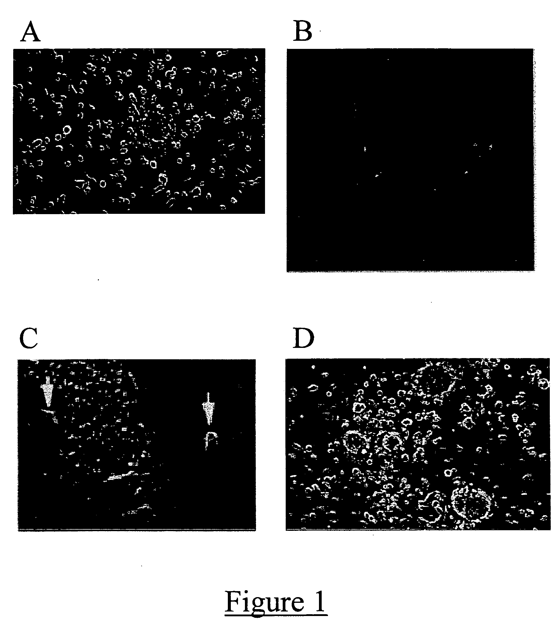
[0039] Whole bone marrow was harvested from the femurs of adult Fisher rats between 16 and 24 weeks of age. Cultures were plated on poly-D-lysine coated 24 well plates at a density of 106 cells per well. The cells were cultured in serum-free Dulbecco's modified Eagle medium (DMEM) / F-12 medium supplemented with B27 (obtained from Gibco BRL; Gaithersburg, Md.), 20 ng / ml FGF-2 and 20 ng / ml EGF (both available from Sigma Chemical Co.; St. Louis, Mo.; hereinafter “Sigma”), along with penicillin and streptomycin (both available from Omega Scientific, Inc.; Tarzana, Calif.).

[0040] After four days in culture, numerous floating spheres of between about 10 to about 100 cells were distinctly visible separate from an underlying adherent monolayer (FIG. 1A). These spheres were collected and sub-cultured separately (FIG. 1B). The cellular aggregates continued to expand and the rate of proliferation remained stable even after multiple disassoci...

example 2

Gene Transfer into Neural Progenitor Cells Utilizing Replication-Deficient Adenoviral Vectors

[0043] Type 5 replication-deficient adenoviral vectors bearing either the reporter gene for β-galactosidase or the gene for the cytokine IL-12 were used to infect neural progenitor cells in vitro. 24 hours following infection, successful gene transfer was confirmed using X-gal staining (X-gal Staining Assay Kit available from Gene Therapy Systems, Inc.; San Diego, Calif.) for α-galactosidase-bearing adenoviral-infected progenitor cells, and an IL-12 Enzyme Linked Immunosorbent Assay (“ELISA” kit available from BD Pharmingen; San Diego, Calif.) for IL-12 gene-bearing adenovirus-infected progenitor cells.

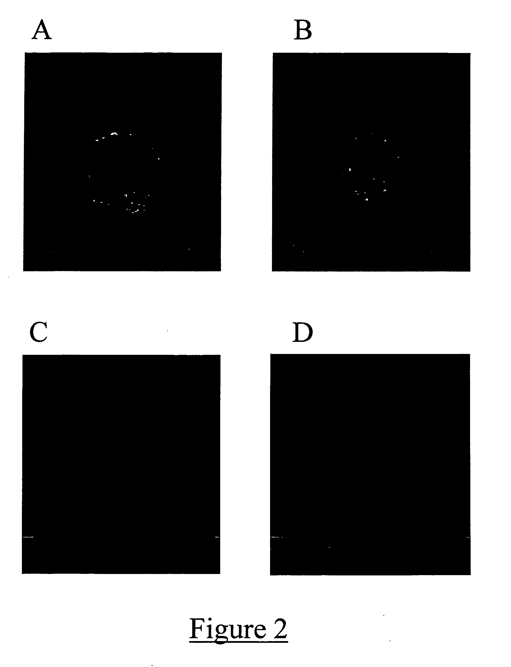
[0044] Successful gene transfer of β-galactosidase was confirmed by positive staining for the X-gal and β-galactosidase-generated blue precipitate in the β-galactosidase-bearing adenovirus-infected progenitor cells (FIG. 5). Successful gene transfer of IL-12 was confirmed by the positive pho...

example 3

Gene Transfer into Neural Progenitor Cells Utilizing a Double-Mutated Herpes Simplex Virus Type I

[0045] A herpes simplex type I virus deleted for the genes encoding the latency activated transcript (LAT) and gamma 34.5 genes (virus denoted DM33) was utilized. The virus contained the gene for GFP under the control of the powerful LAT promoter, and was therefore able to confer constitutive expression of GFP into any successfully infected cell. This vector was used to infect neural progenitor cells in vitro. 72 hours after infection, successful gene transfer was confirmed by viewing GFP expression under a fluorescent light microscope (FIG. 6).

[0046] GFP expression was visible in neural progenitor cells 72 hours following infection with DM33. This confirmed the ability to successfully utilize herpes simplex type I for gene transfer to neural progenitor cells.


PUM

PropertyMeasurementUnit
timeaaaaaaaaaa
massaaaaaaaaaa
volumeaaaaaaaaaa

Abstract

A method is described for generating a clinically significant volume of neural progenitor cells from whole bone marrow. A mass of bone marrow cells may be grown in a culture supplemented with fibroblast growth factor-2 (FGF-2) and epidermal growth factor (EGF). Further methods of the present invention are directed to utilizing the neural progenitor cells cultured in this fashion in the treatment of various neuropathological conditions, and in targeting delivery of cells transfected with a particular gene to diseased or damaged tissue.

Description

[0001] This application claims the benefit of priority under 35 U.S.C. § 119(e) of provisional application Ser. No. 60 / 334,957, filed Oct. 25, 2001, the contents of which are hereby incorporated by reference.FIELD OF THE INVENTION [0002] Embodiments of the present invention are directed to a method for generating a clinically substantial volume of neural progenitor cells from mammalian whole bone marrow. Further embodiments of the present invention are directed to the treatment of neurological disorders using neural progenitor cells cultured in this fashion. BACKGROUND OF THE INVENTION [0003] Nearly every cell in an animal's body, from neural to blood to bone, owes its existence to a stem cell. A stem cell is commonly defined as a cell that (i) is capable of renewing itself; and (ii) can give rise to more than one type of cell (that is, a differentiated cell) through asymmetric cell division. F. M. Watt and B. L. M. Hogan, “Out of Eden: Stem Cells and Their Niches,”Science, 284, 142...

Claims


Application Information

Patent Timeline
Patent Type & Authority Applications(United States)
IPC IPC(8): A61K45/00A61K38/18C12N5/08A61K35/12A61K35/30A61K38/00A61K48/00A61P9/00A61P9/10A61P25/00A61P25/28A61P35/00C12N5/0797
CPCA61K35/12A61K38/00A61K48/00C12N5/0623C12N2506/1353C12N2501/115C12N2506/11C12N2510/00C12N2501/11A61P25/00A61P25/28A61P35/00A61P43/00A61P9/00A61P9/10
Inventor YU, JOHNKABOS, PETEREHTESHAM, MONEEB
Owner CEDARS SINAI MEDICAL CENT
Why Eureka
Try Eureka