Vascular image processing

a technology of image processing and vascular data, applied in the field of vascular data processing, can solve the problems of difficult combining slices into a complete 3d data set, x-ray angiography, visible inner lumen of a diseased vessel,

Inactive Publication Date: 2006-02-16
SHINA SYST
View PDF8 Cites 305 Cited by
  • Summary
  • Abstract
  • Description
  • Claims
  • Application Information

AI Technical Summary

Benefits of technology

[0057] In an exemplary embodiment of the invention, ...

Problems solved by technology

However, combining the slices into a complete 3D data set is not easy.
One limitation of x-ray angiography is that only the inner lumen of a diseased vessel is visible.
Also, the cause of narrowing is not ascertainable, for example, determining if the plaque is fatty or calcified.
It is not believed that sof...

Method used


Image

  • Vascular image processing
  • Vascular image processing
  • Vascular image processing

Examples


Embodiment Construction

Exemplary Vessel Tree

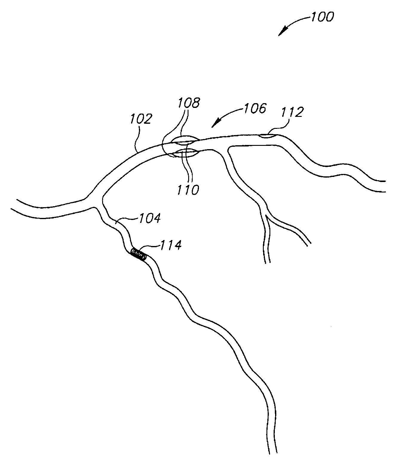
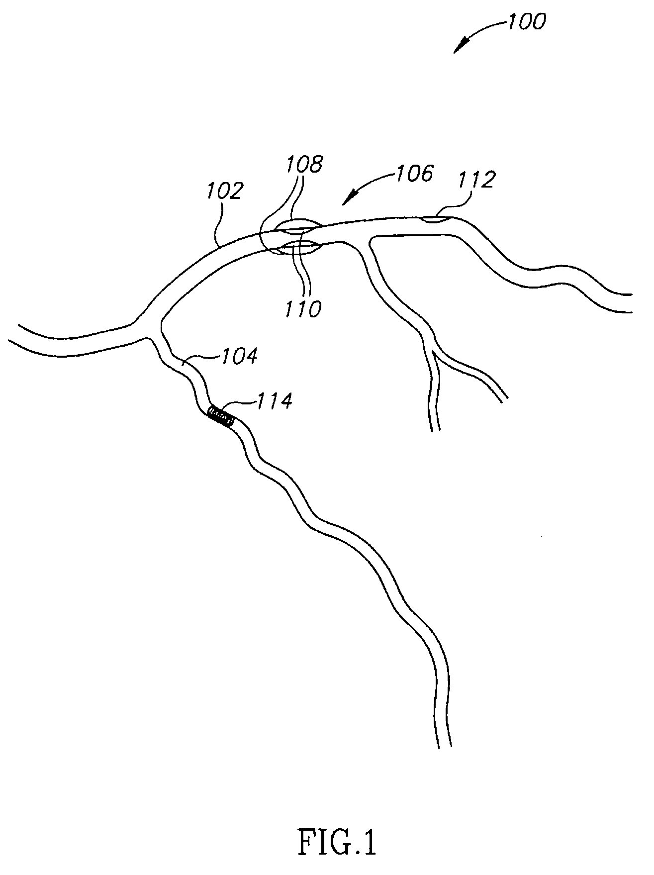
[0080]FIG. 1 shows a vascular tree 100, for example a coronary tree, including various indications such as a stent 114 and regions 106 and 112 narrowed by plaque. As shown the tree includes two main branches 102 and 104 and additional branches (un-numbered) as well.

[0081] Different diagnosis may be required for the different indications, for example, for stent 114, it may be desired to determine a degree of restenosis. For narrowing 108, it may be desirable to determine both a degree of outer remodeling 108 and a degree of lumen narrowing 110. In particular, a type and a dimension of narrowing 110 may be of interest. In an aneurysm (not shown), one or more of outer remodeling, plaque, effect of treatment and / or wall thickness may be of interest. For narrowing 112, which is not visible from the outer vessel diameter (and hence possibly less visible in some 3D imaging methods) the type of narrowing (e.g., soft plaque, calcification, shape, quality or area of ...


PUM


Abstract

A method of estimating a position of a foreign object in the body, comprising: (a) providing a 3D data set of at least one blood vessel; (b) acquiring at least one 2D projection image of the vessel including the object; (c) registering the 2D projection image of the vessel to the 3D data set; and (d) using the registration to estimate a 3D position of said object restricted to be in a blood vessel, according to said 3D data set.

Description

RELATED APPLICATIONS [0001] The present invention claims the benefit under 119(e) of U.S. provisional application 60 / 585,222, filed Jul. 3, 2004, the disclosure of which is incorporated herein by reference.FIELD OF THE INVENTION [0002] The present invention relates to vascular data processing, for example, using CT data to enhance the usability of IVUS data and positioning of catheters on an angiography image. BACKGROUND OF THE INVENTION [0003] A recent development is IVUS (intra vascular ultrasound) in which an IVUS head of a catheter images a slice perpendicular to the vessel axis, using ultrasound imaging. In use, the catheter is pulled back to obtain multiple slices. However, combining the slices into a complete 3D data set is not easy. [0004]“Registration and Analysis of Vascular Images”, Stephen R. Aylward, Julien Jomier, Sue Weeks, Elizabeth Bullitt and “Retrospective registration of x-ray angiogram with MR images by using vessels as intrinsic landmarks”, Journal of Magnetic ...

Claims


Application Information

Patent Timeline
IPC IPC(8): A61B6/00
CPCA61B5/7292A61B6/12A61B6/481A61B6/504A61B8/543A61B6/541A61B8/12A61B8/5238A61B6/5247
Inventor SHINA, YUVAL
Owner SHINA SYST
Why Eureka
Try Eureka